[导读] 编译整理:王小西
Appendiceal serrated lesions and polyps
阑尾锯齿状病变和息肉
Definition
定义
Appendiceal serrated lesions and polyps are mucosal epithelial polyps characterized by a serrated (sawtooth or stellate) architecture of the crypt lumen.
阑尾锯齿状病变和息肉是黏膜上皮息肉,表现为隐窝腔的锯齿状(锯齿状或星状)结构。
ICD-O coding
ICD-O 编码
8213/0 Serrated dysplasia, low grade
8213/2 Serrated dysplasia, high grade
Hyperplastic polyp
Sessile serrated lesion without dysplasia
8213/0 锯齿状异型增生,低级别
8213/2 锯齿状异型增生,高级别
增生性息肉
无蒂锯齿状病变不伴异型增生
ICD-11 coding
ICD-11 编码
2E92.4Y & DB35.0 Other specified benign neoplasm of the large intestine & Hyperplastic polyp of large intestine
2E92.4Y和DB35.0 其他特定的大肠良性肿瘤和大肠增生性息肉
Related terminology
相关术语
Not recommended: diffuse mucosal hyperplasia; serrated adenoma; sessile serrated polyp/adenoma.
不建议:弥漫性黏膜增生;锯齿状腺瘤;无蒂锯齿状息肉/腺瘤。
Subtype(s)
亚型
None
无
Localization
部位
Serrated lesions and polyps can occur throughout the appendix.
锯齿状病变和息肉可发生于整个阑尾。
Clinical features
临床特征
Most serrated lesions and hyperplastic polyps (HPs) are incidental findings detected in appendices removed for other reasons. Large lesions may cause obstruction and lead to appendicitis, potentially complicated by rupture.
大多数锯齿状病变和增生性息肉(HP)是在因其他原因切除的阑尾中检测到的偶然发现。大病变可能引起阻塞,并导致阑尾炎,可能并发破裂。
Epidemiology
流行病学
Appendiceal serrated polyps occur in men and women with about equal frequency. Patients are generally older adults, in their sixth to eighth decade of life, although there is a wide age range {3679,2488}.
阑尾锯齿状息肉在男性和女性中发生的频率大致相同。患者通常是50至80岁的老年人,但年龄范围较广{3679,2488}。
Etiology
病因
Unknown
未知
Pathogenesis
发病机制
HPs and mucosal hyperplasia can be seen in postinflammatory reparative settings, including after an episode of acute appendicitis, in diverticular disease of the appendix, and in interval appendectomy. However, genetic alterations raise the possibility that some of these polyps are neoplastic. KRAS or BRAF mutations are found in some hyperplastic lesions {3679,2488}. KRAS mutations are frequent in serrated polyps of the appendix, particularly in serrated polyps with dysplasia. BRAF mutations are less common but have been found in a subset of serrated polyps {3679,2488}. These studies suggest that KRAS may be more biologically important in the appendix than BRAF, and that the serrated pathway of carcinogenesis may have less relevance in the appendix than in the colon.
增生性息肉和黏膜增生可见于炎症后修复环境中,包括急性阑尾炎发作后、阑尾憩室病患病期间和阑尾切除术后。然而,遗传学改变提示其中一些息肉可能是肿瘤性。在一些增生性病变中发现KRAS或BRAF突变{3679,2488}。KRAS突变常见于阑尾锯齿状息肉,尤其是伴有异型增生的锯齿状息肉。BRAF突变不太常见,但已在一些锯齿状息肉发现{3679,2488}。这些研究表明,KRAS在阑尾中可能比BRAF更具有生物学意义,锯齿状癌变途径在阑尾中的相关性可能低于结肠。
Macroscopic appearance
肉眼外观
Serrated lesions may form a discrete polyp or may circumferentially involve the appendiceal mucosa.
锯齿状病变可能形成离散的息肉,也可能环周累及阑尾黏膜。
Histopathology
组织病理学
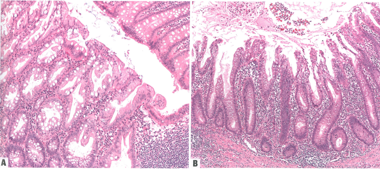
In appendiceal HPs, similar to in colonic HPs, the affected mucosa shows elongated crypts with increased numbers of goblet cells or a mixture of goblet cells and columnar cells with smaller mucin vacuoles (1228,2625). The luminal part of the crypts shows a serrated crypt profile. Cytological atypia is mild at most, generally in the deep crypts, and can be attributed to reactive changes. Cytological dysplasia is not present.
与结肠增生性息肉相似,阑尾增生性息肉的受累黏膜显示隐窝变长伴杯状细胞数量增多,或杯状细胞与柱状细胞混合,杯状细胞的黏液空泡较小(1228,2625)。隐窝的管腔部分显示锯齿状隐窝轮廓。细胞学异型性最多为轻度,一般在深部隐窝,可能归因于反应性改变。不存在细胞学异型增生。
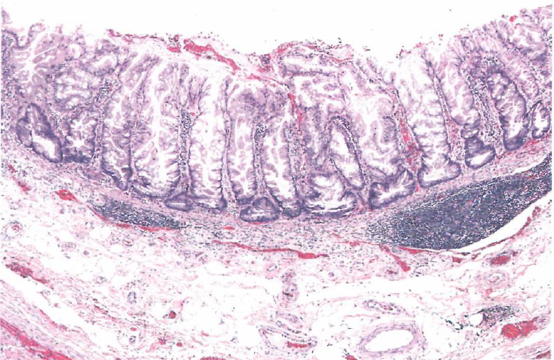
In serrated lesions without dysplasia, the mucosa demonstrates abnormal crypt proliferation with elongated and serrated crypt profiles. Serration and dilation extend to the crypt bases with abnormal shapes including L shapes and inverted T shapes. Varying degrees of villous architecture may be seen. There may be mild cytological atypia with dystrophic goblet cells and possibly more mitotic figures than would be seen in an HP. Luminal mucin may be abundant {292,2776}.
在不伴异型增生的锯齿状病变中,黏膜表现为异常隐窝增殖,隐窝轮廓细长且锯齿状。锯齿状和扩张延伸至隐窝底部,形状异常,包括L形和倒T形。可见到不同程度的绒毛结构。可能存在轻度细胞学异型性伴异型增生的杯状细胞,核分裂象可能多于增生性息肉。管腔黏液可能丰富 {292,2776}。
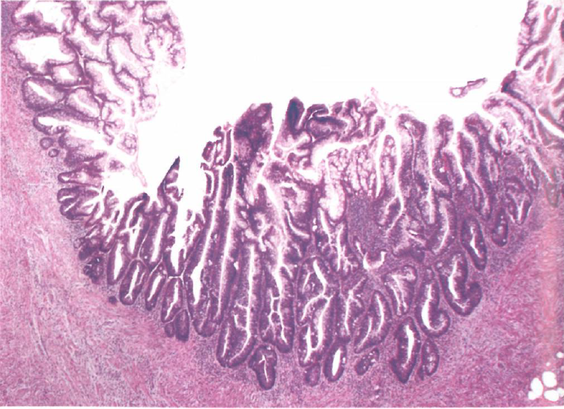
In serrated lesions with dysplasia, dysplasia can take the form of conventional adenoma-like dysplasia, serrated dysplasia, or traditional serrated adenoma-like dysplasia, and multiple morphological patterns of dysplasia may be seen within a single polyp {3679}. The dysplastic component may be well demarcated from non-dysplastic areas of the serrated polyp. Conventional adenoma-like dysplasia usually develops a villous growth pattern with elongated, hyperchromatic nuclei with pseudostratification, increased mitoses, and apoptotic bodies, similar to in colorectal adenomas. Serrated dysplasia maintains the serrated architecture of the crypts, but the crypts are lined by cuboidal to low columnar cells with hyperchromatic enlarged nuclei, reduced cytoplasmic mucin, and increased mitoses. Traditional serrated adenoma-like dysplasia shows complex serration and villous growth, with the villi lined by tall columnar cells with eosinophilic cytoplasm. The nuclei are elongated and mildly hyperchromatic, but the degree of atypia is less than in conventional adenoma-like dysplasia. The villi may have abortive-type crypts along their lateral edges, which has also been described in traditional serrated adenomas in the colorectum. The dysplasia can be classified as low-grade or high-grade.
在伴有异型增生的锯齿状病变中,异型增生可以表现为传统的腺瘤样异型增生、锯齿状异型增生或传统的锯齿状腺瘤样异型增生,并且在单个息肉中可以看到异型增生的多种形态学模式{3679}。异型增生成分可与锯齿状息肉的非异型增生区域分界清楚。传统的腺瘤样异型增生通常形成绒毛状生长模式,细胞核拉长深染,并有核假复层、核分裂增加和凋亡小体,与结直肠腺瘤相似。锯齿状异型增生主要形成隐窝的锯齿状结构,但隐窝内衬立方形至低柱状细胞,伴核深染增大、细胞质黏液减少和核分裂增加。传统的锯齿状腺瘤样异型增生表现为复杂的锯齿状和绒毛状生长,绒毛被覆高柱状细胞,胞质嗜酸性。细胞核拉长,轻度深染,但异型程度不及传统的腺瘤样异型增生。绒毛沿其侧缘可有流产型隐窝,在结直肠传统锯齿状腺瘤中也有描述。异型增生可分为低级别或高级别。
Like serrated lesions and polyps, low-grade appendiceal mucinous neoplasms (LAMNs) can show serrated architecture, but they usually show areas with long filiform villi without serration. In contrast to LAMN, which often results in loss of the lamina propria and muscularis mucosae as well as mural fibrosis, serrated polyps retain the normal architecture of the appendix and typically have an intact muscularis mucosae and lamina propria. In addition, tumours with pushing invasion and submucosal fibrosis or dissemination to the peritoneum as pseudomyxoma peritonei are probably LAMNs and should not be classified as serrated polyps. Some tumours are heterogeneous, with areas resembling serrated polyp and other areas resembling LAMN; in such cases, the lesion is best classified as LAMN.
与锯齿状病变和息肉一样,低级别阑尾黏液性肿瘤(LAMN)可显示锯齿状结构,但通常显示无锯齿状结构的长丝状绒毛区域。LAMN通常导致固有层和黏膜肌层缺失以及管壁纤维化,相反,锯齿状息肉保留了阑尾的正常结构,通常具有完整的黏膜肌层和固有层。此外,具有推挤浸润和黏膜下纤维化或腹膜播散成为腹膜假黏液瘤的肿瘤可能是LAMN,不应归类为锯齿状息肉。一些肿瘤具有异质性,有些区域类似锯齿状息肉,其他区域类似LAMN;在这种情况下,病变最好归类为LAMN。
Cytology
细胞学
Not clinically relevant
无临床相关性
Diagnostic molecular pathology
诊断分子病理学
Not clinically relevant
无临床相关性
Essential and desirable diagnostic criteria
基本和理想的诊断标准
See Table 5.01.
见表5.01。
Staging (TNM)
分期(TNM)
Not clinically relevant
无临床相关性
Prognosis and prediction
预后和预测
Not clinically relevant
无临床相关性

Fig. 5.01 Hyperplastic polyp of the appendix. A High-power view of a hyperplastic polyp (bottom) and adjacent normal crypts (right); the crypts in the hyperplastic polyp show serration of the luminal aspect of the crypt and no dysplasia. B The mucosa does not protrude above the surface, but the crypts show serrated profiles towards their luminal aspects.
Fig. 5.01 阑尾增生性息肉。A 高倍显示增生性息肉(底部)和相邻的正常隐窝(右侧);增生性息肉中的隐窝显示隐窝腔面有锯齿状,无异型增生。B 黏膜未突出于表面上方,但隐窝显示朝向其管腔方面的锯齿状轮廓。

Fig. 5.02 Appendiceal sessile serrated lesion without dysplasia. The crypts are serrated and the base of the crypts shows abnormal architecture, with crypt dilatation and inverted T shapes.
Fig. 5.02 不伴异型增生的阑尾无蒂锯齿状病变。隐窝为锯齿状,隐窝底部结构异常,伴有隐窝扩张和倒T形。

Fig. 5.03 Appendiceal sessile serrated lesion with adenoma-like dysplasia. In the centre of the field, the polyp shows hyperchromatic pseudostratified nuclei consistent with dysplasia, whereas at the sides of the image, the polyp is serrated but lacks cytological dysplasia.
Fig. 5.03 阑尾无蒂锯齿状病变伴腺瘤样异型增生。在视野中心,息肉显示深染的假复层细胞核,符合异型增生,而在图像两侧,息肉呈锯齿状,但无细胞学异型增生。


来源:
Digestive System Tumours: WHO Classification of Tumours (Medicine)
WHO Classification of Tumours Editorial Board
第5版本
World Health Organization
ISBN-13: 978-9283244998
ISBN-10: 9283244990
仅供学习交流,不得用于其他任何途径。如有侵权,请联系删除。


共0条评论