[导读] 以下病例来源于《Gastrointestinal Cancer Atlas for Endoscopic Therapy》Rikiya Fujita, Hiroshi Takahashi (Eds.),如有兴趣可购买原书阅读。
【CASE1】


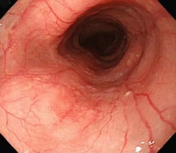
Routine observation: The lesion exhibited a mildly reddish depression without granular irregularity, but mucosal extension was preserved. Disruption of branch-like blood vessels was visualized (arrows)
Iodine staining: Note the multiple unstained areas. The lesion was seen as an 8-mm area that was unstained with iodine and located exactly at the reddish mucosa


NBI: The lesion was visualized as a brownish area
Magnifying NBI: Type V vascular pattern is revealed


Pathological fi ndings: Squamous cell carcinoma, 0-IIc, T1a-EP, ly0, v0, HM0, VM0
Diagnostic Points:
The lesion was easily recognized by observations with NBI and iodine staining.
Mucosal redness with disrupted blood vessels is a fi nding that should never be overlooked by routine observation.
【CASE2】


Routine observation: A 6-mm area with mild redness was observed (arrows). There was no mucosal irregularity. In terms of invasion depth, T1a-EP cancer was suspected
Iodine staining: There was an area unstained with iodine that corresponded to the site of a reddish depression seen by routine observation. The site was positive for the pink color sign


NBI: The lesion was visualized as a brownish area (arrows) that corresponded to the site of a reddish depression seen by routine observation
Magnifying NBI: Type V1 vascular pattern was revealed


Pathological fi ndings: Squamous cell carcinoma, 0-IIb, T1a-EP, ly0, v0, HM0, VM0
Diagnostic Points:
Metachronous multiple lesions were detected during the follow-up after endoscopic resection of a superfi cial-type esophageal cancer. It is important to pay close attention to the presence of metachronous multiple lesions. The fi nding of a positive pink color sign can help diagnose a small lesion.
【CASE3】


Routine observation: An area unstained with iodine extended longitudinally from the lower esophagus (arrows). Reexamination after administration of a proton pump inhibitor (PPI) revealed a mildly reddish area 6 mm in diameter at the same region
Iodine staining: An area unstained with iodine (arrows) was observed at exactly the same site of a reddish depression observed by routine observation. The site was positive for the pink color sign


NBI: The lesion was visualized as a brownish area corresponding to the site of the reddish depression detected by routine observation
Magnifying NBI: Note that blood vessels are thin and extended owing to infl ammation (arrows); a V3 vascular pattern was observed in part. Invasion depth TIb-MM to SM1 was suspected


Pathological findings: Squamous cell carcinoma, 0-IIc, T1b-SM (127 μm), ly0, v0, HM0, VM0
Diagnostic Points:
Background infl ammation was severe, and the margin was unclear. After PPI administration,inflammation subsided and the margin became clearer. A positive pink color sign would help diagnose the margin of the lesion.


共0条评论