[导读] 以上病例来源于《Gastrointestinal Cancer Atlas for Endoscopic Therapy
》如有兴趣可购买原书阅读。
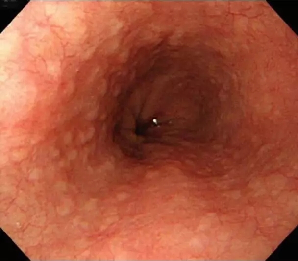
如果是你,先看看这两个食管有没有问题?有问题的地方在哪里?


可能有点难度.....
【CASE4】


Routine observation: A mildly reddish area of about 8 mm was seen (arrows). Given the absence of evident granular changes at the redness, an invasion depth T1a-EP cancer was suspected
白光下可见一约8mm轻度发红的病变,鉴于未见颗粒感改变,故考虑上皮内肿瘤可能性大。
Iodine staining: An area unstained with iodine was observed at the site of the redness seen by routine observation
碘染后病对应区域呈负染区。


NBI: The lesion was visualized as a brownish area (arrows) at the site of the redness detected by routine observation
NBI下观察病变对应区域呈茶褐色。
Magnifying NBI: Type VI vascular pattern was revealed
放大观察可见血管大部呈井上分型的V1型(个人认为也可见V2型)


Pathological findings: Squamous cell carcinoma, 0-IIc, T1a-LPM, ly0, v0, HM0, VM0
病理提示为固有层鳞癌,切缘阴性,淋巴血管阴性。
Diagnostic Points:
Iodine-unstained areas were scattered, but a positive pink color sign was recognized only at this site, which would help lead to the diagnosis.
诊断观察要点:在碘染多发分散的情况下,请注意粉红征的变化。
【CASE5】


 Routine observation: The lesion (arrows) was difficult to detect by routine observation
白光观察很难发现的一病变。(0-IIb,5-mm)
Iodine staining: The lesion was seen as an area unstained with iodine and with a positive pink color sign
碘染后呈负染区,粉红征阳性。


NBI: The lesion was visualized as a brownish area (arrows)
Magnifying NBI: Type V1 vascular pattern was revealed
NBI下呈茶褐色改变,放大观察IPCL大部分呈井上分型V1型改变,少量V2。


Pathological findings: Squamous cell carcinoma, 0-IIc, T1a-LPM, ly0, v0, HM0, VM0
病理提示为固有层鳞癌,切缘阴性,淋巴血管阴性。
Diagnostic Points:
The iodine-unstained area with a positive pink color sign and a type V vascular
pattern are suggestive of malignancy.
诊断观察要点:碘染呈负染,粉红征阳性,V型血管均提示肿瘤性病变。
【CASE6】


Routine observation: There was a reddish depression protruding from the squamous-columnar junction (SCJ) to the squamous cell epithelium in the shape of a tongue. Although lower esophageal palisade vessels were unclear, the SCJ appeared irregular; and the presence of short-segment Barrett’s esophagus (SSBE) was suspected
位于SCJ的浅凹陷发红的舌形病变拟为SSBE,表面结构不规则,表面血管不清。
Indigo carmine staining: Depression of the lesion became clearer. There was no surface irregularity at the depression
靛胭脂染色后病变清晰,表面结构基本规则。


Magnifying NBI: Reticular vascular pattern was observed at the depression, strongly suggesting malignancy
Magnifying NBI: A change in mucosal structure at the margin of the depression was revealed, which better demarcated the boundary
网格状血管形态高度提示肿瘤性病变,边界线清晰存在。

Pathohistological findings: Adenocarcinoma (tub 1), M, ly0, v0, HM0, VM0
病理提示粘膜内高分化管状腺癌,切缘阴性,血管淋巴阴性。
Diagnostic Point:
It is necessary to observe carefully the reddish mucosa at the SCJ. In particular, when SSBE is present, detecting a change in mucosal structure and abnormal blood vessels by magnifying NBI would help with the diagnosis.
观察诊断要点:仔细观察SCJ附近的发红区域,当发现 SSBE时,进一步放大观察表面结构及血管形态至关重要。


 Routine observation: The lesion (arrows) was difficult to detect by routine observation


共0条评论