[导读] 来源:
The Practice of Surgical Pathology:A Beginner’s Guide to the Diagnostic Process
外科病理学实践:诊断过程的初学者指南
Diana Weedman Molavi, MD, PhD
Sinai Hospital, Baltimore, Maryland
ISBN: 978-0-387-74485-8 e
第2章 解剖病理学中的描述性术语(Descriptive Terms in Anatomic Pathology)
Central to effective learning in pathology is the ability to speak the language. This chapter covers the approach to defining and describing an unknown tumor or lesion and defines histologic terms commonly used in pathology.
学习病理学最有效方法是语言表达能力。本章涵盖了定义和描述未知肿瘤或病变的方法,并定义了病理学常用的组织学术语。
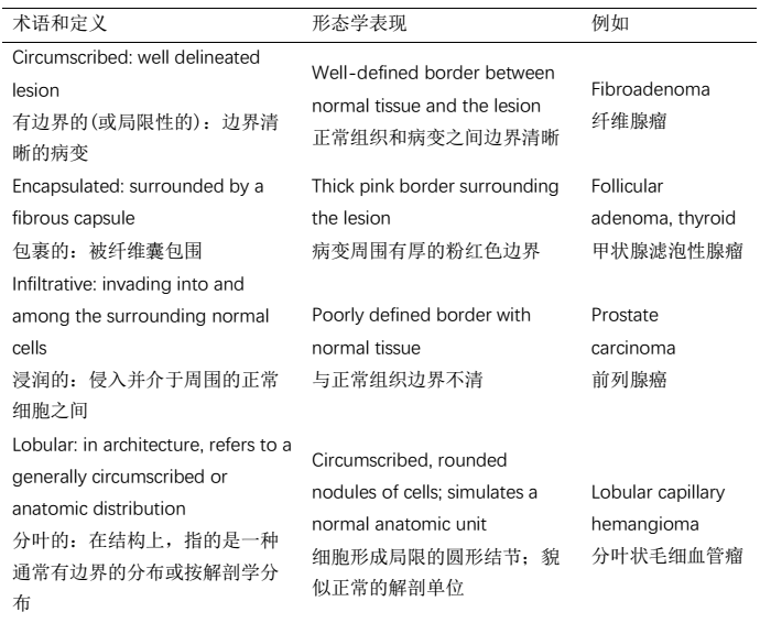
2.1与周围正常组织的界面(Interface With the Surrounding Normal Tissue)

2.2细胞量(从低到高)和核分裂率(Cellularity (Low to High) and Mitotic Rate)
Note the cellularity (by cellularity we often mean how blue it is, or how densely packed the nuclei are). Cellularity may be described as hypercellular or just cellular or as hypocellular/ paucicellular. Also look for mitoses on high power. High mitotic rate may be an indicator of malignancy. Atypical mitoses (tripolar) are convincing indicators of malignancy. Estimate how many mitoses are seen per high power field (40×).
细胞量是指细胞丰富程度(我们通常所说的细胞量是指它有看上去有多蓝,或细胞核有多密集)。细胞量可描述为细胞量丰富、不加描述的细胞量或细胞量稀少/少细胞性。同时在高倍镜下寻找核分裂象。高核分裂率(或核分裂活跃)可能是恶性肿瘤的一个指标。非典型核分裂象(三极核分裂象)是恶性肿瘤令人信服的指标。估计每个高倍视野(40×)能看到多少核分裂象。
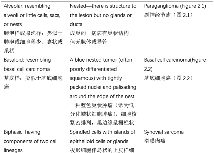
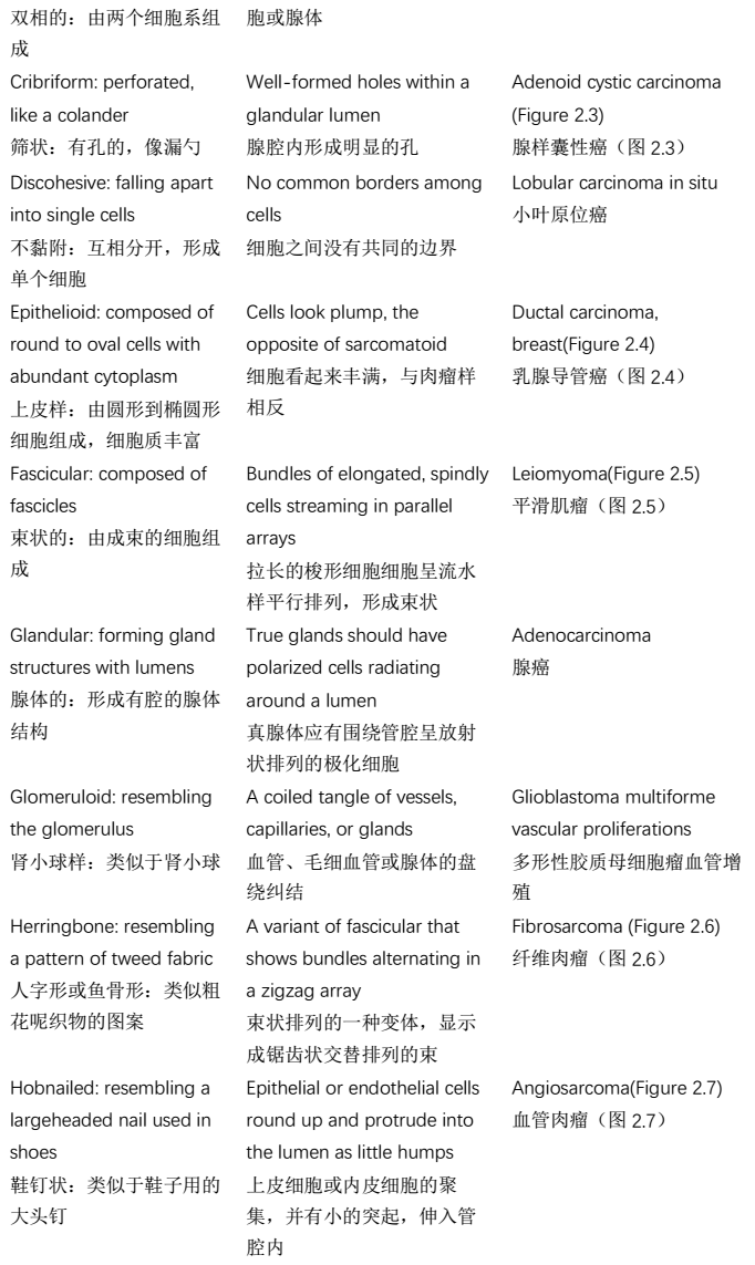
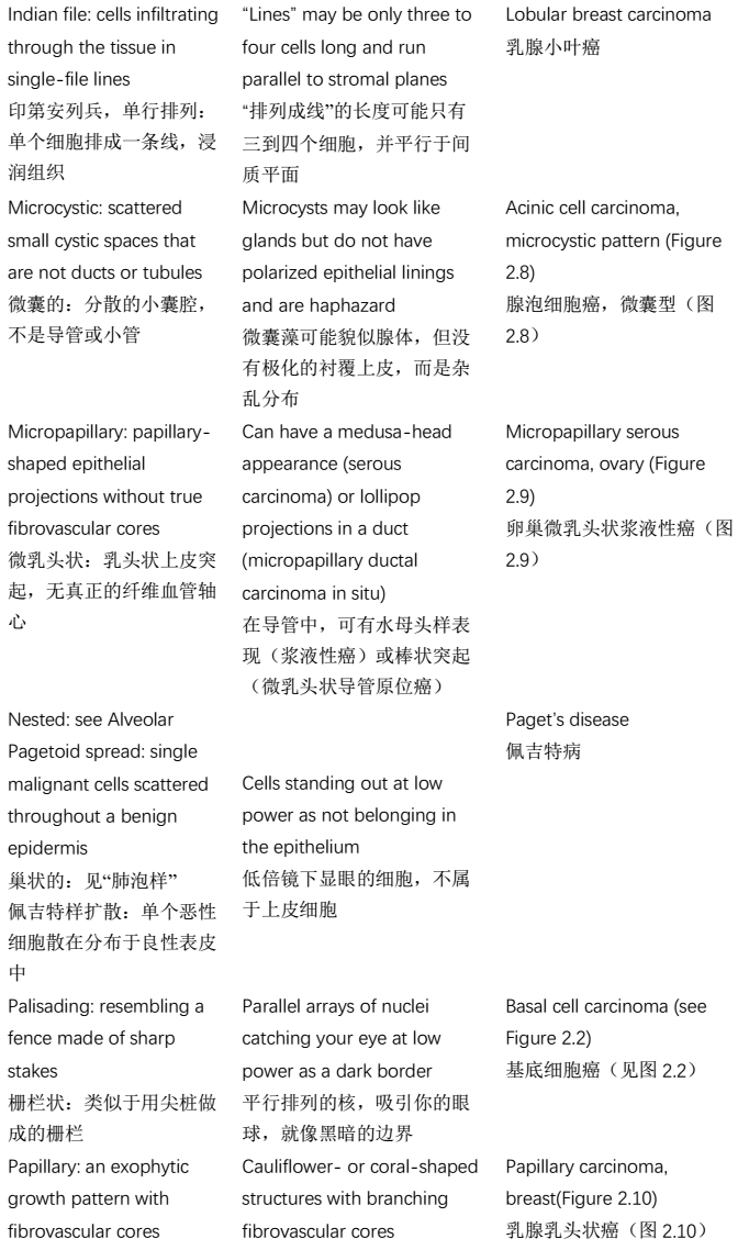
2.3结构模式(Architectural Pattern)
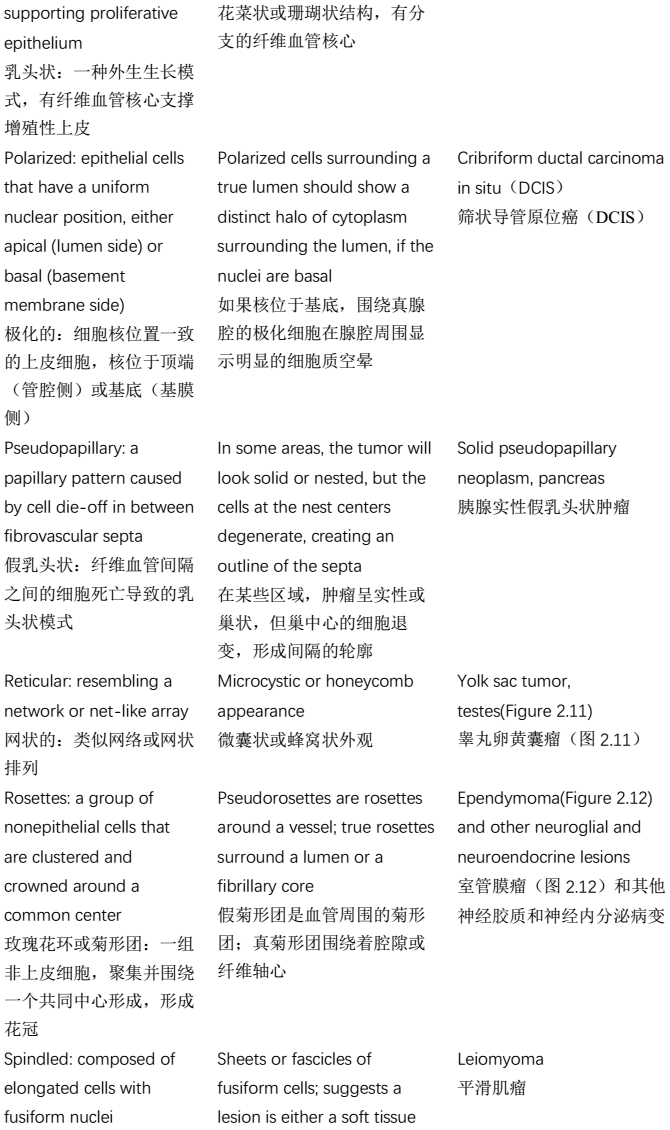
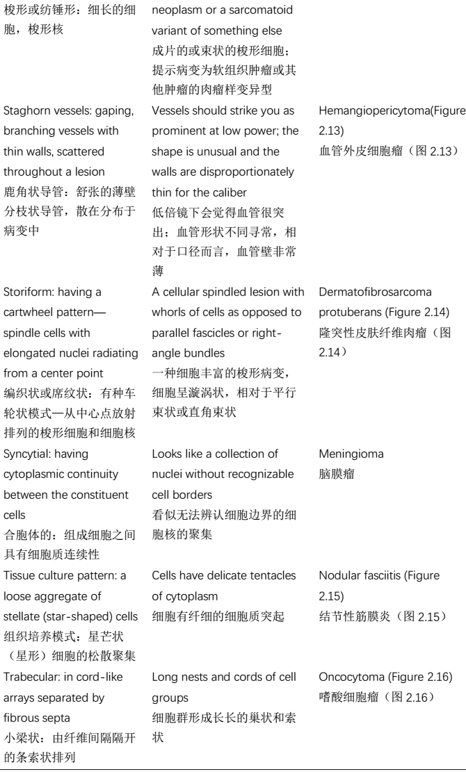
2.4有无坏死(Presence or Absence of Necrosis)

2.5细胞形状和大小,细胞质(Cell Shape and Size, Cytoplasm)

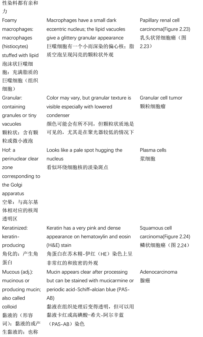
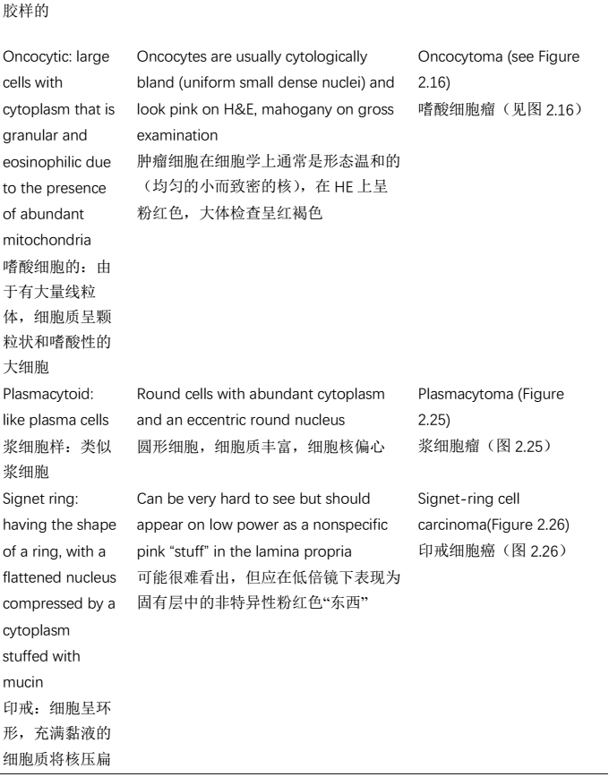
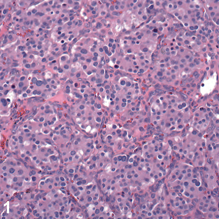
Figure 2.1. Alveolar pattern, paraganglioma.
图2.1.腺泡状结构,副神经节瘤。
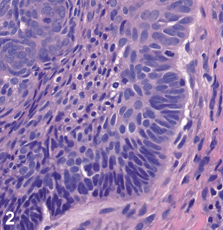
Figure 2.2. Basaloid pattern and palisading, basal cell carcinoma.
图2.2.基底样结构和栅栏状排列,基底细胞癌。
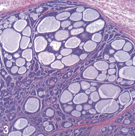
Figure 2.3. Cribriform pattern, adenoid cystic carcinoma.
图2.3.筛状结构,腺样囊性癌。
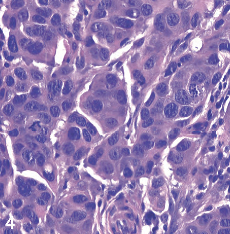
Figure 2.4. Epithelioid cells, breast carcinoma.
图2.4.上皮样细胞,乳腺癌。
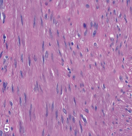
Figure 2.5. Fascicular pattern, leiomyoma.
图2.5.束状结构,平滑肌瘤。

Figure 2.6. Herringbone pattern, fibrosarcoma.
图2.6.人字形,纤维肉瘤。
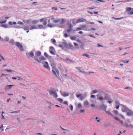
Figure 2.7. Hobnailed cells, angiosarcoma.
图2.7.鞋钉状细胞,血管肉瘤。
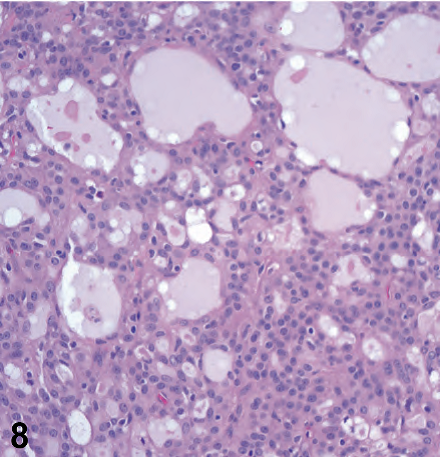
Figure 2.8. Microcystic pattern, acinic cell carcinoma.
图2.8.微囊结构,腺泡细胞癌。
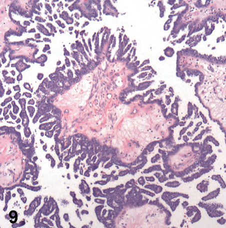
Figure 2.9. Micropapillary architecture, micropapillary serous carcinoma of the ovary carcinoma.
图2.9.微乳头状结构,卵巢癌的微乳头状浆液性癌。
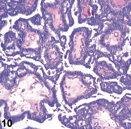
Figure 2.10. Papillary architecture, papillary carcinoma of breast.
图2.10.乳头状结构,乳腺乳头状癌。

Figure 2.11. Reticular pattern, yolk sac tumor of testis.
图2.11.网状结构,睾丸卵黄囊瘤。
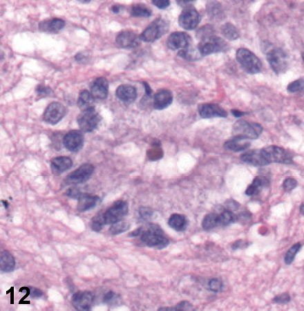
Figure 2.12. Rosette, ependymoma.
图2.12.菊形团,室管膜瘤。
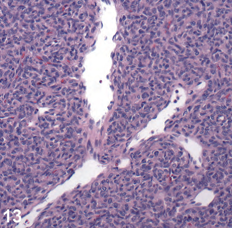
Figure 2.13. Staghorn vessels, hemangiopericytoma.
图2.13.鹿角状血管,血管外皮细胞瘤。
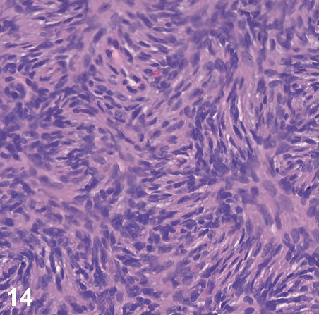
Figure 2.14. Storiform pattern, dermatofibrosarcoma protuberans.
图2.14.席纹状结构,隆突性皮肤纤维肉瘤。
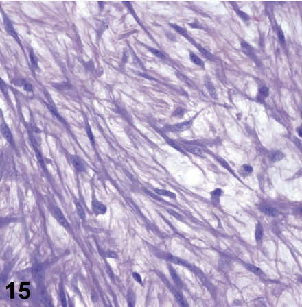
Figure 2.15. Tissue culture cells, nodular fasciitis.
图2.15.组织培养样细胞,结节性筋膜炎。
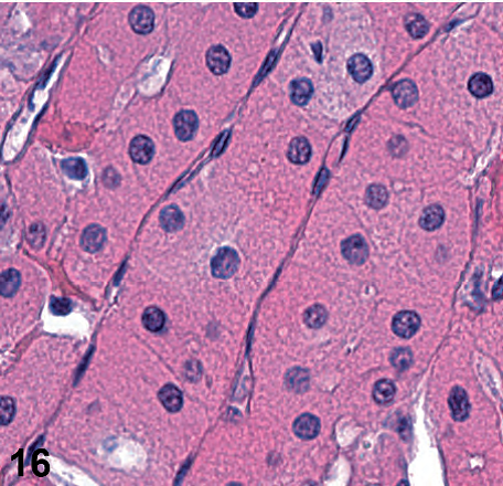
Figure 2.16. Trabecular pattern and oncocytes, oncocytoma.
图2.16.小梁结构和嗜酸细胞,嗜酸细胞瘤。

Figure 2.17. Coagulative necrosis, ischemic bowel.
图2.17.凝固性坏死,肠缺血。
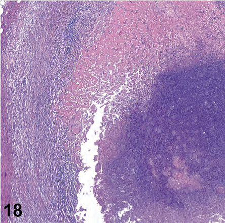
Figure 2.18. Caseating necrosis in a granuloma, tuberculosis.
图2.18.肉芽肿中的干酪样坏死,结核。
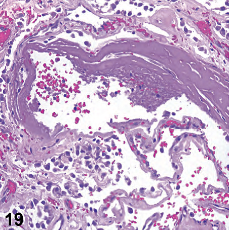
Figure 2.19. Fibrinoid necrosis, pulmonary vessel.
图2.19.纤维素样坏死,肺血管。
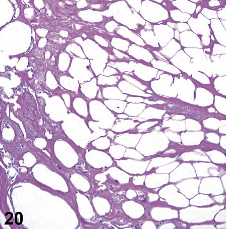
Figure 2.20. Fat necrosis, breast.
图2.20.乳房脂肪坏死。
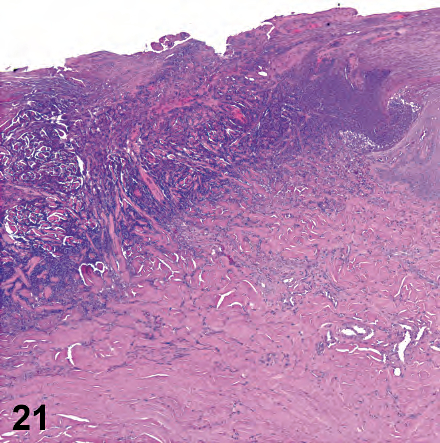
Figure 2.21. Gangrenous necrosis, toe wound.
图2.21.坏疽性坏死,脚趾伤口。
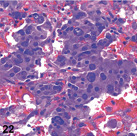
Figure 2.22. Amphophilic cytoplasm, pheochromocytoma.
图2.22.双染性细胞质,嗜铬细胞瘤。
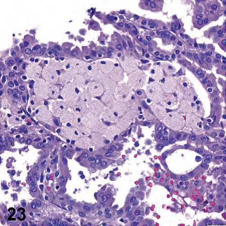
Figure 2.23. Foamy macrophages, papillary renal cell carcinoma.
图2.23.泡沫状巨噬细胞,乳头状肾细胞癌。
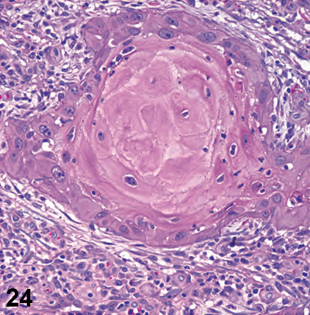
Figure 2.24. Keratin, squamous cell carcinoma.
图2.24.角蛋白,鳞状细胞癌。
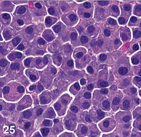
Figure 2.25. Plasmacytoid, plasmacytoma.
图2.25.浆细胞样,浆细胞瘤。
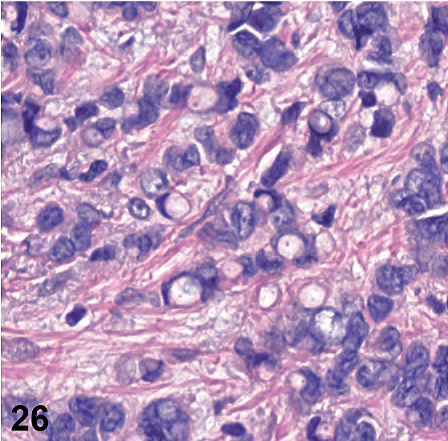
Figure 2.26. Signet-ring cells, breast carcinoma.
图2.26.印戒细胞,乳腺癌。

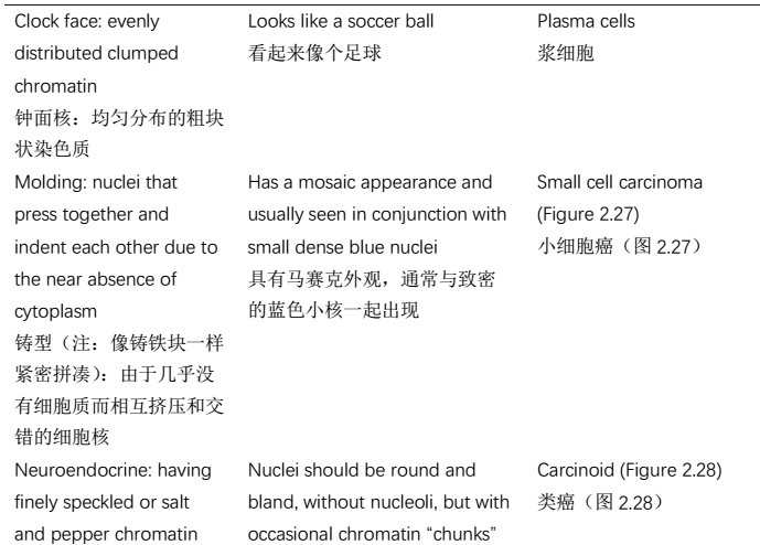
Figure 2.27. Nuclear molding, small cell carcinoma.
图2.27.核铸型,小细胞癌。
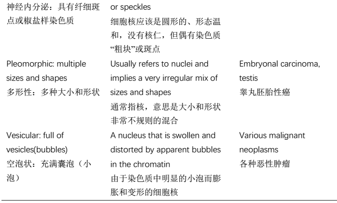
2.6细胞核,或简称核(Nucleus)
2.7核仁(Nucleolus)

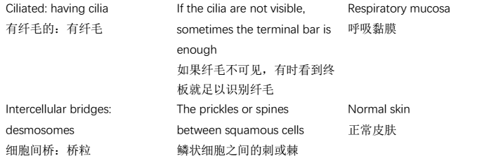
2.8细胞膜(Cell Membrane)
2.9病变的间质,如果有(Stroma of Lesion, If Present)


2.10其他非细胞成分(Other Noncellular Entities)

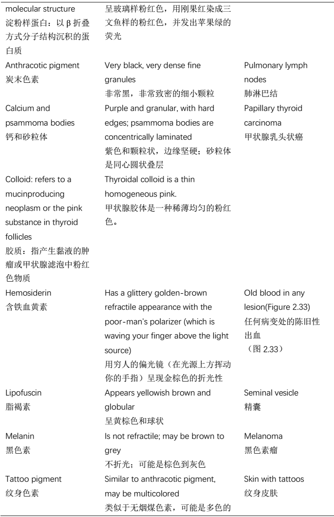
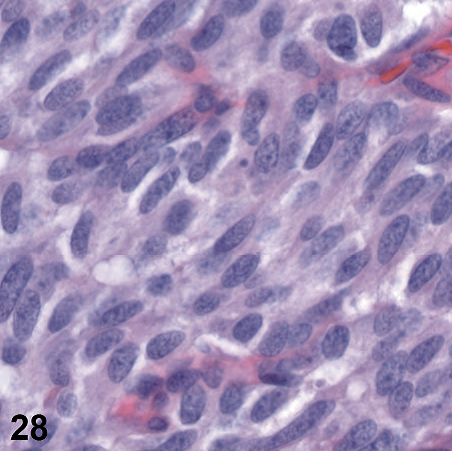
Figure 2.28. Neuroendocrine nuclei, carcinoid tumor.
图2.28.神经内分泌细胞核,类癌。
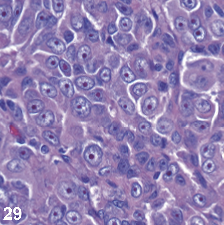
Figure 2.29. Cherry-red nucleolus, melanoma.
图2.29.樱桃红色核仁,黑色素瘤。
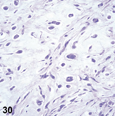
Figure 2.30. Myxoid stroma, myxoid myxofibrosarcoma.
图2.30.黏液样间质,黏液样黏液纤维肉瘤。
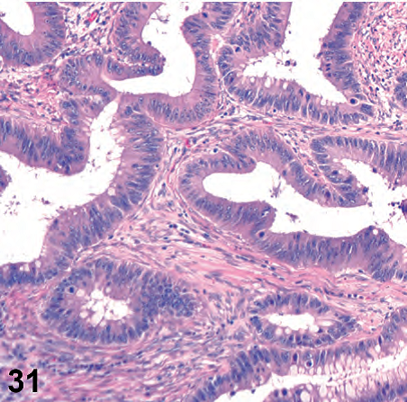
Figure 2.31. Desmoplastic stroma, colon cancer.
图2.31.促结缔组织增生性间质,结肠癌。
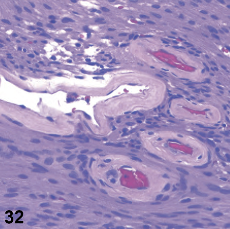
Figure 2.32. Hyaline deposits, vessels in schwannoma.
图2.32.神经鞘瘤血管中的透明沉积物。
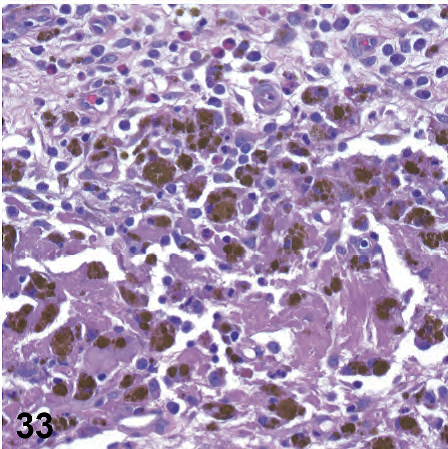
Figure 2.33. Hemosiderin, nasal polyp.
图2.33.含铁血黄素,鼻息肉。
来源:
The Practice of Surgical Pathology:A Beginner’s Guide to the Diagnostic Process
外科病理学实践:诊断过程的初学者指南
Diana Weedman Molavi, MD, PhD
Sinai Hospital, Baltimore, Maryland
ISBN: 978-0-387-74485-8 e-ISBN: 978-0-387-74486-5
Library of Congress Control Number: 2007932936
© 2008 Springer Science+Business Media, LLC
仅供学习交流,不得用于其他任何途径。如有侵权,请联系删除。


共0条评论