外科病理学实践:诊断过程的初学者指南 | 第28章 软组织和骨 下
骨骼肌/横纹肌肿瘤(Skeletal Muscle Tumors)
Tumors of skeletal muscle are uncommon, and, as a terminal cell type with no stem cells or regenerative activity (like neurons), they are mainly seen in children or young adults (Table 28.6). They all get the rhabdo- prefix and should all stain with actin and desmin, plus special skeletal muscle markers myogenin and MyoD1. Other unrelated tumors in other sites sometimes are described as “rhabdoid;” remember that the –oid suffix means “looks like, but is not.” Therefore, the rhabdoid meningioma or rhabdoid tumor is not of muscle origin but simply displays similar cell shape and appearance.
骨骼肌/横纹肌肿瘤少见,作为一种没有干细胞或再生活性(如神经元)的终末细胞类型,主要见于儿童或年轻人(表28.6)。它们都有rhabdo-(杆状的-)前缀,并且都应该染actin和desmin染色,加上特异性骨骼肌标记物myogenin和MyoD1。其他部位的其他不相关肿瘤有时称为“横纹肌样”;记住–oid后缀的意思是“看起来像,但不是”。因此,横纹肌样脑膜瘤或横纹肌样瘤不是肌肉来源,只是显示类似的细胞形状和外观。
表28.6 骨骼肌/横纹肌肿瘤。
Table 28.6. Skeletal muscle neoplasms.
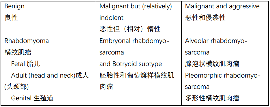
The rhabdomyoma is a rare neoplasm of primitive skeletal muscle cells. The fetal rhabdomyoma generally occurs in children, typically in the head and neck, and resembles the embryonal rhabdomyosarcoma but without the atypia and mitoses. In adult men, the adult rhabdomyoma usually arises in the head and neck. In this variant, the cells are rhabdoid in shape, with small peripheral nuclei, and pink clumps of myofilaments in the cytoplasm, not unlike mature muscle cut in cross-section. The third variant, genital, occurs in adult women. This variant is predominantly composed of strap cells (elongated pink cells with cytoplasmic cross-striations), again without atypia or mitoses.
横纹肌瘤是一种罕见的原始骨肌细胞肿瘤。胎儿横纹肌瘤通常发生在儿童,通常发生在头颈部,与胚胎性横纹肌肉瘤相似,但没有异型性和核分裂。成人横纹肌瘤通常发生在头部和颈部。在这种变异型中,细胞呈横纹肌样,周围有小的细胞核,细胞质中有粉红色的肌丝束,与成熟肌肉的横切面相似。第三种变异型,生殖器,发生在成年女性身上。这种变异型主要由条带细胞(带细胞质横纹的细长粉红色细胞)组成,同样没有异型性或核分裂。
Rhabdomyosarcoma is the most common sarcoma of children and is rare in adults. Remember that any high-grade sarcoma can acquire some rhabdo- differentiation, however. Pure rhabdomyosarcomas can be grouped into three subtypes: embryonal, alveolar, and pleomorphic. The pleomorphic type is found in adults.
横纹肌肉瘤是儿童最常见的肉瘤,成人罕见。记住,任何高级别肉瘤都可以获得一些横纹肌分化。纯横纹肌肉瘤可分为三种亚型:胚胎性、腺泡状和多形性。多形性见于成人。
Embryonal rhabdomyosarcoma comprises about 80% of cases and has a significantly better outcome than the alveolar type. It is composed of sheets of rhabdomyoblasts (plump and eosinophilic with large eccentric nuclei), nonspecific spindled cells, or strap cells (Figure 28.15). The botryoid subtype of embryonal rhabdomyosarcoma refers to tumors occurring in a mucosal site, such as the genital tract.
胚胎性约占病例的80%,其预后明显优于腺泡状。它由成片的横纹肌母细胞(丰满、嗜酸性、核大偏心)、非特异性梭形细胞或条带状细胞组成(图28.15)。胚胎性横纹肌肉瘤的葡萄簇样亚型是指发生在黏膜部位的肿瘤,如生殖道。
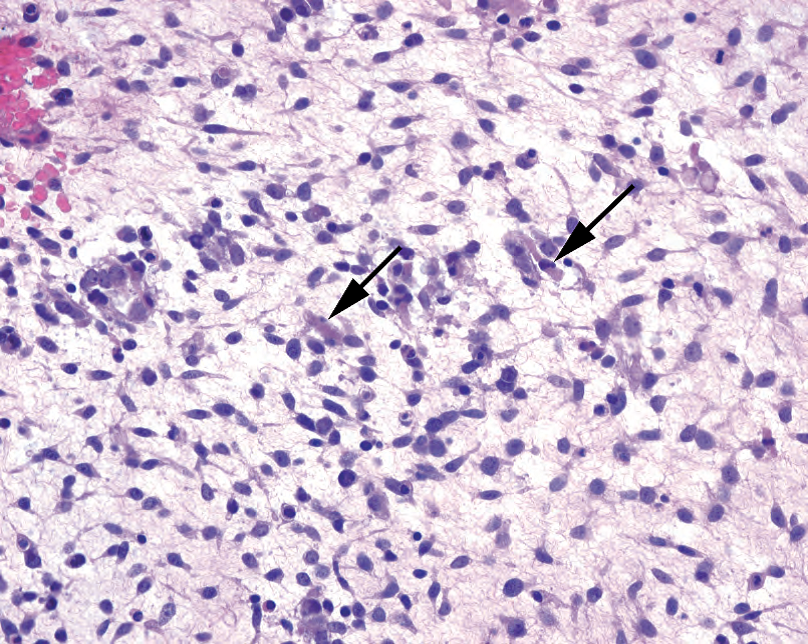
Figure 28.15. Embryonal rhabdomyosarcoma, botryoid type. The background is gelatinous, and the small spindle cells are nonspecific in appearance. Occasional strap cells are visible, with cytoplasmic muscle fibers (arrows).
图28.15胚胎性横纹肌肉瘤。背景为黏液样,小梭形细胞的形态学无特异性。偶见带状细胞,有细胞质肌纤维(箭)。
Alveolar rhabdomyosarcoma is a very aggressive variant and equals “unfavorable histology.” In this type, fibrous septa divide the tumor into packets, much like renal cell carcinoma, but the discohesive cells tend to fall apart in the middle of the packets (Figure 28.16). The solid variant may be indistinguishable from the small round blue cell tumors. As it turns out, the cytogenetic findings are different in embryonal and alveolar types. As the prognosis is so different, many centers are beginning to routinely do molecular tests on these tumors.
腺泡状横纹肌肉瘤是一种侵袭性很强的变异型,等于“不良的组织学”。在这种类型中,纤维间隔将肿瘤分成小巢,很像肾细胞癌,但不粘附的细胞在细胞巢中间往往是散开的(图28.16)。实性变异型可能与小圆蓝细胞肿瘤难以区分。事实证明,胚胎性和腺泡状的细胞遗传学结果是不同的。由于预后如此不同,许多中心开始对这些肿瘤进行常规的分子检测。
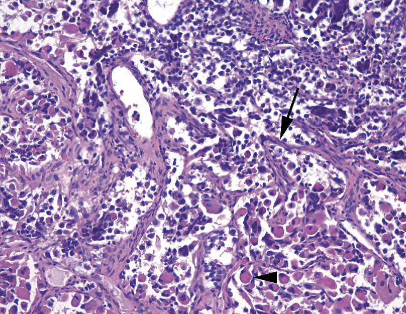
Figure 28.16. Alveolar rhabdomyosarcoma. The alveolar pattern is outlined by fibrovascular septa (arrow), and the tumor cells tend to fall out of the centers of their nests. This example shows prominent rhabdomyoblast differentiation (arrowhead), with large cells full of dense pink cytoplasm and eccentric nuclei. Other specimens may show only small round blue cell phenotype.
图28.16 腺泡状横纹肌肉瘤。纤维血管间隔(箭)勾画出腺泡状模式,肿瘤细胞倾向于从细胞巢中心脱落。本例显示明显的横纹肌母细胞分化(箭头),大细胞充满致密的粉红色细胞质,核偏心。其他病例可能仅显示小圆蓝细胞表型。
外周神经/神经外胚层肿瘤(Peripheral Nerve/Neuroectodermal Tumors)
Nerves, as the axonal processes of terminally differentiated neurons around the spinal cord, do not actually form tumors. However, the cells associated with the nerve sheath do commonly produce neoplasms, including schwannoma and neurofibroma. Other tumors of neuroectodermal origin are included here as well.
神经,作为脊髓周围的终末分化神经元的轴突,实际上并不形成肿瘤。然而,与神经鞘相关的细胞通常会产生肿瘤,包括神经鞘瘤和神经纤维瘤。这里也包括其他起源于神经外胚层的肿瘤。
The only lesion that could be called reactive, in this group, is the traumatic neuroma. This is a disorganized tangle of nerve endings, including Schwann cells, perineurium, and axons, that may be found at the location of prior surgery or trauma. On the slide, it looks like a large, frayed nerve.
这组病变中,唯一可以称为反应性病变的是创伤性神经瘤。这是一种紊乱的神经末梢缠结,包括雪旺细胞、神经束膜和轴突,可见于先前的手术或创伤部位。在切片片上,它看起来像一条巨大的、磨损的神经。
The benign peripheral nerve sheath tumors include the schwannoma and the neurofibroma (Table 28.7). Both of these lesions are S100 positive. Both can undergo malignant transformation into the malignant peripheral nerve sheath tumor, although this is much less common in the schwannoma. However, the nerve sheath lesions, just like the pleomorphic lipoma, may occasionally acquire bizarre cytology that does not indicate malignancy. This degenerative atypia is called ancient change.
良性外周神经鞘肿瘤包括神经鞘瘤和神经纤维瘤(表28.7)。这两种病变均为S100阳性。两者都可以恶性转化为恶性的外周神经鞘肿瘤,尽管这种情况在神经鞘瘤中很少见。然而,神经鞘病变,就像多形性脂肪瘤一样,偶尔也会出现非恶性的奇异细胞学。这种退化的非典型称为“古老改变"(退变)。
The schwannoma is an encapsulated lesion that arises from a peripheral nerve and can therefore be found anywhere in the body. It usually shows alternating hypercellular (Antoni A) and myxoid (Antoni B) areas, as well as characteristic parallel arrays of palisading cells called Verocay bodies (Figure 28.17). The cells themselves have euchromatic, fusiform nuclei that stream in parallel within a pink fibrillary background. Thick-walled, hyalinized vessels are typical, which look as though they have a layer of amyloid replacing the vessel wall.
神经鞘瘤是一种由外周神经引起的包裹性病变,因此可以在身体的任何地方发现。它通常显示交替的富细胞(Antoni A)区和黏液样少细胞(Antoni B)区,以及称为Verocay小体的栅栏状细胞的特征性平行阵列(图28.17)。细胞本身有正常染色的梭形细胞核,在粉红色的纤丝状背景中平行流动。厚壁玻璃样血管是典型表现,看起来好像有一层淀粉样蛋白取代了血管壁。
The cellular variant of schwannoma is still benign but can get quite hypercellular, and mitotically active (up to 10 mitoses per 10 hpf). The capsule and hyaline vessels should help to point you toward schwannoma. Foamy macrophages are common within this tumor.
神经鞘瘤的富细胞变异型仍然是良性的,但可有相当多的细胞,并且核分裂活跃(多达10/10HPF)。包膜和透明血管有助于辨认神经鞘瘤。这种肿瘤中常见泡沫状巨噬细胞。
The neurofibroma, in contrast, is an unencapsulated lesion that may appear as a nodule, a poorly circumscribed tumor, or a plexiform (“bag of worms”) tangle. It is pale to pink at low power, with a myxoid background and thin curly tendrils of collagen between the cells (Figure 28.18). The nuclei are pale, thin, and slightly undulating, as in a normal nerve, and there should be no mitoses. Unlike in the schwannoma, special stains may reveal axons trapped within the lesion.
相反,神经纤维瘤是一种无包膜的病变,可表现为结节、边界不清的肿瘤或丛状(“虫袋”)缠结。低倍镜下呈淡染至粉红色,细胞间有黏液样背景和细薄卷曲的胶原丝(图28.18)。细胞核淡染,细长,轻微起伏,就像正常神经的核,应该没有核分裂。与神经鞘瘤不同的是,特殊染色可显示病变内的轴突。
表28.7 神经相关肿瘤。
Table 28.7. Nerve-related neoplams.
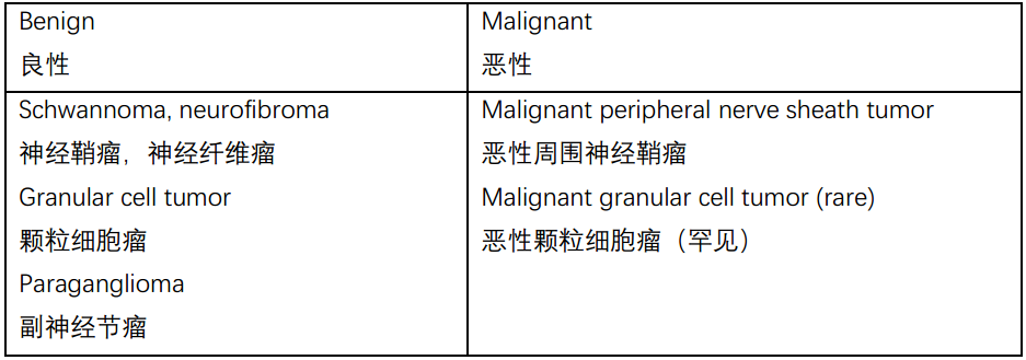
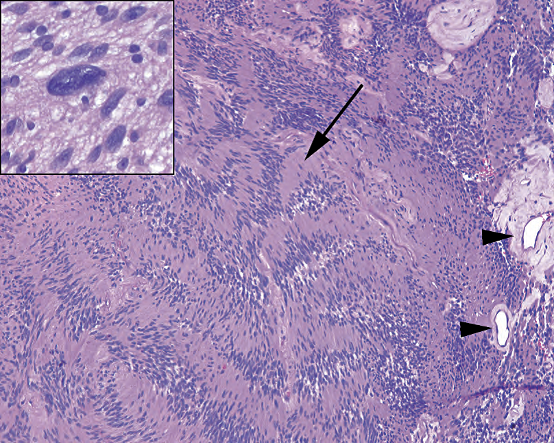
Figure 28.17. Schwannoma. In this spindle cell neoplasm, the long tapered nuclei tend to clump together and form arrays called Verocay bodies (arrow). Hyalinized vessels (arrowheads) are common. Inset: Occasional large atypical cells indicate ancient change, not malignancy.
图28.17 神经鞘瘤。在这例梭形细胞肿瘤中,核细长,一端渐细,核趋向于聚集在一起,排列成Verocay小体(箭)。常见玻璃样血管(箭头)。插图:偶见非典型大细胞,提示退变,而不是恶性肿瘤。
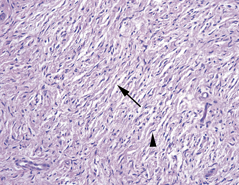
Figure 28.18. Neurofibroma. The nuclei tend to be thin and wavy (arrowhead), much like in a normal nerve. The tumor is usually paucicellular, with a myxoid background and delicate curly strands of wispy collagen (arrow).
图28.18 神经纤维瘤。细胞核往往细长、波浪状(箭头),很像正常神经的核。肿瘤通常是少细胞的,有黏液样背景和纤细卷曲的胶原丝(箭头)。
The malignant peripheral nerve sheath tumor is usually a high-grade sarcoma and often takes the morphology of the fibrosarcoma (Figure 28.19). It may retain some nerve sheath features, such as the wavy nuclei, nuclear palisading, or hyalinized vessels, but tends to lose most of its S100 reactivity. Mitoses should be present, unlike in a neurofibroma.
恶性周围神经鞘瘤通常是一种高级别肉瘤,通常具有纤维肉瘤的形态(图28.19)。它可能保留一些神经鞘特征,如波状核、核栅栏状排列或透明血管,但大部分肿瘤细胞往往失去S100反应性。应当存在核分裂,这与神经纤维瘤不同。
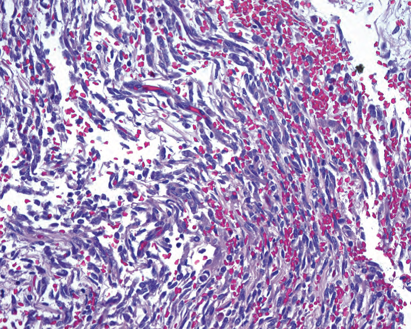
Figure 28.19. Malignant peripheral nerve sheath tumor. Although the malignant peripheral nerve sheath tumor sometimes resembles a fibrosarcoma, in this example it is more reminiscent of a neurofibroma, which was probably the origin in this case. There is a myxoid background and wavy collagen, but the cells are much more hyperchromatic and atypical than in a neurofibroma.
图28.19 恶性周围神经鞘瘤。虽然恶性周围神经鞘瘤有时类似纤维肉瘤,但本例更像神经纤维瘤,提示本例可能起源于神经纤维瘤。有黏液样背景和波浪状胶原,但细胞比神经纤维瘤更为深染,非典型更严重。
The granular cell tumor is a benign tumor that shows neural differentiation but that resembles a collection of foamy macrophages. It is often associated with striking pseudoepitheliomatous hyperplasia in mucosal sites. These epithelial changes may be mistaken for squamous cell carcinoma if the subtle underlying diagnostic granular cells are overlooked.
颗粒细胞瘤是一种良性肿瘤,显示神经分化,但含有大量泡沫巨噬细胞。它常伴随黏膜部位显著的假上皮瘤样增生。如果忽略表皮下方细微的诊断性颗粒细胞,这些上皮变化可能会误认为是鳞状细胞癌。
The paraganglioma is actually a neuroendocrine tumor but is included here as it is s ometimes presents as a soft tissue mass. It is a (usually) benign tumor with neuroendocrine-type nuclei, arranged in an alveolar pattern (Figure 28.20).
副神经节瘤实际上是一种神经内分泌肿瘤,但由于它有时表现为软组织肿块,因此也在这里提及。它(通常)是一种良性肿瘤,具有神经内分泌型细胞核,呈腺泡状排列(图28.20)。
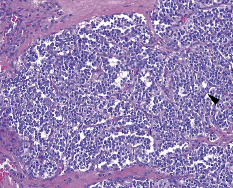
Figure 28.20. Paraganglioma. Fibrovascular septa (arrowhead) divide the neoplasm into small balls of cells (the “zellballen” pattern). The cells have small, perfectly round nuclei with neuroendocrine chromatin. Despite the paraganglioma’s classification as an extraadrenal pheochromocytoma, it resembles the carcinoid tumor more closely than the pheochromocytoma.
图28.20 副神经节瘤。纤维血管间隔(箭头)将肿瘤分割成小球(细胞球)。这些细胞有小而圆的细胞核和神经内分泌型染色质。尽管副神经节瘤被分类为肾上腺外嗜铬细胞瘤,但它比嗜铬细胞瘤更像类癌。
血管肿瘤(Vascular Tumors)
Reactive lesions of capillaries are very common, as an inherent part of the healing process is the formation of new vasculature. Granulation tissue, which fills in a defect in the body tissues, has very prominent capillaries with large endothelial cells (see Figure 3.2 in Chapter 3). The capillaries of granulation tissue are plump and round, with at least two cell layers (endothelium and pericytes), and may be crowded but do not appear interconnected. Neoplastic vessels, on the other hand, are often lacking the pericyte component and typically form anastomotic channels and slit-like spaces with sharp angular profiles. Extravasated blood cells are common in vascular neoplasms. The immunohistochemical markers for the vascular tumors are CD31 and CD34.
毛细血管的反应性病变很常见,因为愈合过程中固有的一部分是新生血管的形成。填补身体组织缺损的肉芽组织有非常明显的毛细血管和大的内皮细胞(见第3章图3.2)。肉芽组织的毛细血管肿胀变圆,至少有两层细胞(内皮细胞和血管周细胞),血管可能拥挤,但不会相互连接成网。另一方面,肿瘤性血管通常缺乏周细胞成分,通常形成吻合血管,血管腔呈裂隙状并有尖角外形。血管肿瘤常见血细胞外渗。血管肿瘤的免疫组化标记物为CD31和CD34。
Papillary endothelial hyperplasia is a pattern of organizing thrombus that may occur within a vessel or hematoma. It may be seen incidentally in a surgical specimen or represent a symptomatic small mass by itself, in which case it is called a Masson’s tumor. It is composed of tiny fibrin papillae covered by thin endothelium (Figure 28.21).
乳头状内皮增生是机化血栓的一种模式,可能发生在血管内或血肿内。它可能偶见于外科标本,也可能本身就是一个有症状的小肿块,后者称为Masson瘤。它由细小的纤维素乳头组成,被覆薄的内皮(图28.21)。
A hemangioma is a benign neoplasm of vascular elements, and there are many subtypes, including the common capillary hemangioma, the cavernous hemangioma, and the juvenile hemangioma (Table 28.8). The hemangiomas generally have round, nonbranching vessels, although they may be very crowded or dilated, and the capillaries are surrounded by a pericyte layer. The pyogenic granuloma, once thought to be a reactive lesion, may in fact be a true neoplasm and is now called lobular capillary hemangioma. It is a circumscribed mass of capillaries with associated inflammation and ulceration. This lobular (circumscribed with rounded contours) appearance is characteristic of benign vascular lesions in general.
血管瘤是血管成分的良性肿瘤,有许多亚型,包括常见的毛细血管瘤、海绵状血管瘤和幼年性血管瘤(表28.8)。血管瘤通常有圆形、无分支的血管,血管可能非常拥挤或扩张,毛细血管周围有一层周细胞层。化脓性肉芽肿,曾经认为是一种反应性病变,实际上可能是一种真正的肿瘤,现称为分叶状毛细血管瘤。它是一个由边界的毛细血管肿块,伴有炎症和溃疡。总体上,这种分叶状结构(边界清楚的圆形轮廓)是良性血管病变的特征。
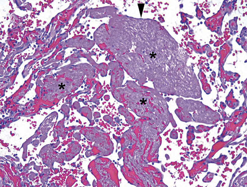
Figure 28.21. Papillary endothelial hyperplasia. Fingers of fibrin and red blood cells (asterisks), not true fibrovascular cores, are lined by bland endothelial cells (arrowhead).
图28.21 乳头状内皮增生。纤维素和红细胞(星号)的形成的手指状结构,不是真正的纤维血管轴心,被覆形态学温和的内皮细胞(箭头)排列。
表28.8 血管肿瘤。
Table 28.8. Vascular neoplams.
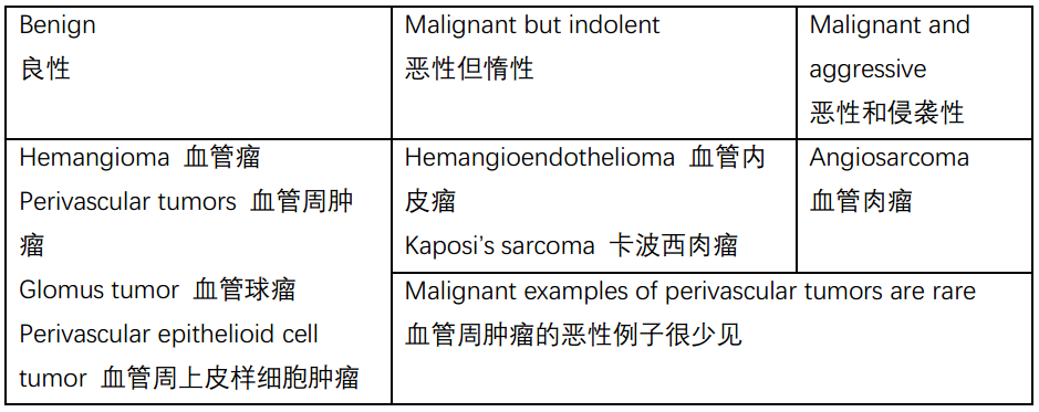
For every category of endothelial lesions there is an epithelioid variant, in which the endothelial cells acquire a lot of cytoplasm, becoming plump and epithelial-looking, often with cytoplasmic lumina that are their attempts at vessels. These variants are challenging because they may not look particularly vascular. Negative epithelial markers (cytokeratins, EMA) are helpful, but unfortunately some epithelioid vascular neoplasms may express some keratins.
内皮病变的每个类别都有一种上皮样变异型,其内皮细胞获得大量细胞质,变得丰满并呈现上皮外观,通常具有细胞质空腔,这是它们在试图形成血管。这些变异型具有挑战性,因为它们看起来可能不太像血管。阴性上皮标记物(CK,EMA)有助于区分,但不幸的是,一些上皮样血管肿瘤可能表达某些角蛋白。
The indolent malignant lesions of endothelium are called hemangioendothelioma. The epithelioid hemangioendothelioma is a sclerosing lesion with cords of vacuolated cells, some of which may contain red blood cells within the vacuoles, a diagnostic feature (Figure 28.22). It can be very difficult to distinguish from carcinoma without stains.
内皮的惰性恶性病变称为血管内皮瘤。上皮样血管内皮瘤是一种硬化性病变,空泡状细胞形成条索,其中一些空泡内可能含有红细胞,这是一种诊断特征(图28.22)。如果没有免疫染色,很难与癌区分。
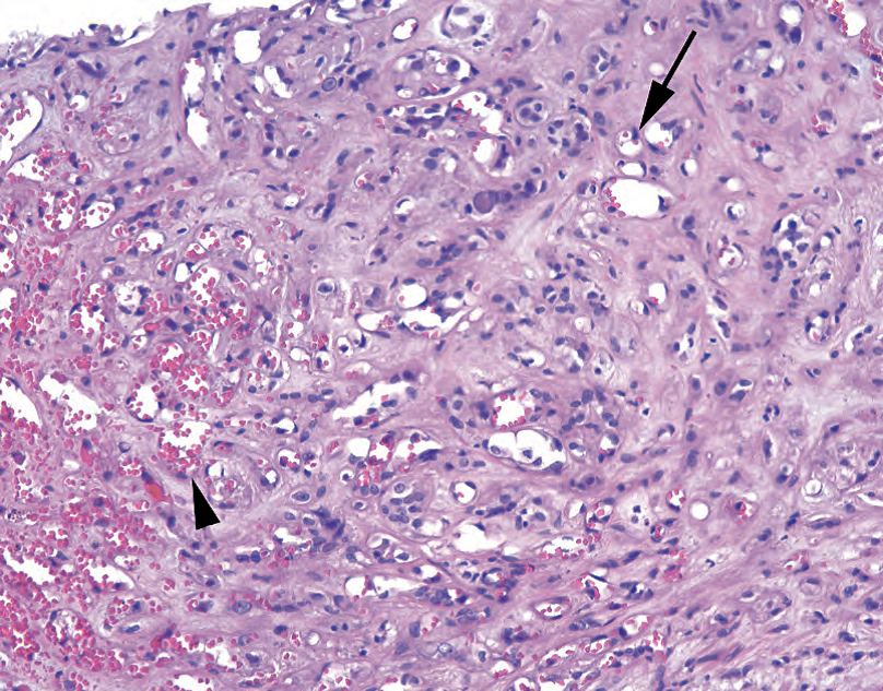
Figure 28.22. Epithelioid hemangioendothelioma. A rare but distinctive tumor, the epithelioid hemangioendothelioma is characterized by a dense fibrotic or sclerotic background, with small capillary spaces (arrowhead) and single cells with intracytoplasmic lumens complete with red blood cells (arrow).
图28.22 上皮样血管内皮瘤。这是一种罕见但独特的肿瘤,其特征是致密的纤维化或硬化背景,有小的毛细血管腔(箭头),单个细胞有胞质内空腔,空腔含有完整的红细胞(箭)。
Kaposi’s sarcoma, a virally induced (human herpesvirus type 8) low-grade sarcoma seen primarily in patients with HIV, has several stages and appearances, ranging from the most subtle of slit-like spaces in the dermis (see Chapter 27) to a dense spindle cell lesion. Because of the many variants, and a considerable array of “Kaposiform” mimickers, the differential diagnosis is beyond the scope of this chapter.
卡波西肉瘤是一种病毒诱导的(人类疱疹病毒8型,HHV8)低级别肉瘤,主要见于HIV患者,有几个阶段和表现,从真皮内最细微的狭缝状裂隙(见第27章)到致密的梭形细胞病变。由于有许多变异型,以及大量的“卡波西样”类似病变,鉴别诊断超出了本章的范围。
Angiosarcoma is the high-grade endothelial tumor, and it too has many variants. It can occur in organs, such as the liver or breast, especially after exposure to toxins or radiation. However, it can also arise in soft tissues de novo. Lymphedema is a recognized risk factor. Angiosarcoma classically shows branching, anastomotic irregular spaces with bulbous atypical cells lining the spaces (the hobnail pattern; Figure 28.23). Pericytes are typically absent, and at the periphery the tumor infiltrates into the surrounding tissue. This infiltrative border is very helpful in identifying malignant lesions.
血管肉瘤是一种高级别的内皮细胞肿瘤,它也有许多变异型。它可以发生在器官,如肝脏或乳房,尤其是暴露于毒素或辐射后。然而,它也可以在软组织从头发生。淋巴水肿是公认的风险因素。血管肉瘤典型表现为分支、吻合的不规则腔隙,腔隙内衬覆球状非典型细胞(鞋钉状;图28.23)。周细胞通常不存在,肿瘤的边缘浸润到周围组织中。这种浸润性边缘对鉴别恶性病变很有帮助。
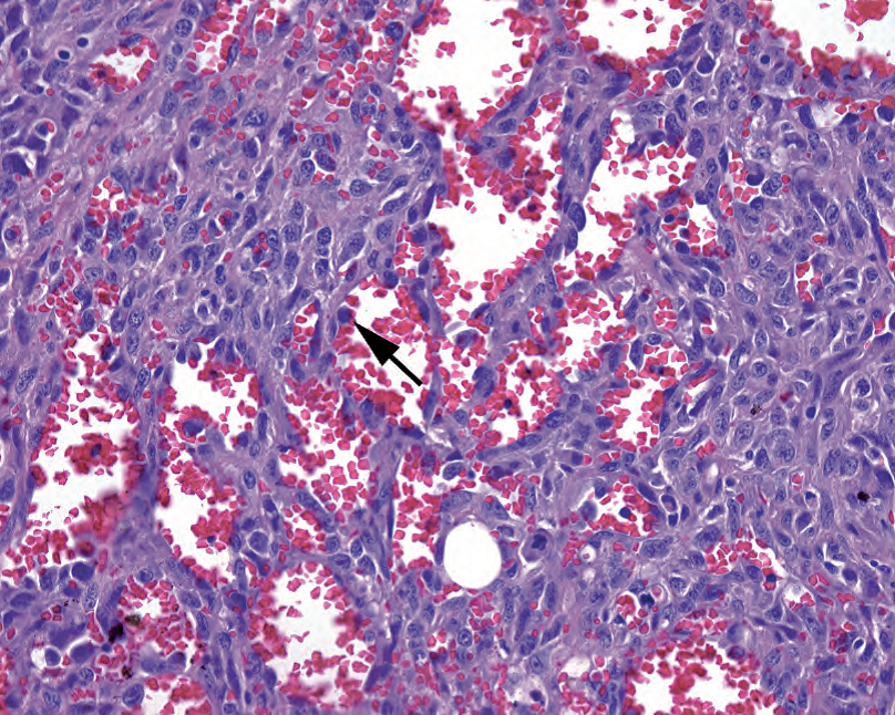
Figure 28.23. Angiosarcoma. In some areas the tumor cells have begun to grow as a solid sheet, but there are still vascular spaces visible, and full of blood. Large and hyperchromatic malignant cells protrude into the lumina (arrow) in a hobnail pattern. The tumor cells also show prominent nucleoli.
图28.23 血管肉瘤。部分区域,肿瘤细胞已开始生长成为一个实性成片,但仍有可见的血管腔,并充满血液。大而深染的恶性细胞以鞋钉状突向管腔内(箭)。肿瘤细胞也有明显的核仁。
Naturally, there is an epithelioid variant of angiosarcoma. The epithelioid angiosarcoma may look like a generic “very bad tumor” composed of sheets of plump cells with large nuclei and prominent nucleoli, having almost no vascular differentiation. This sort of tumor may be identified only after a large battery of stains.
自然地,血管肉瘤有一种上皮样变异型。上皮样血管肉瘤可能看起来像一种普通的“很坏的肿瘤”,由成片的丰满的细胞组成,细胞核大,核仁明显,几乎没有血管分化。这种肿瘤只有经过大量免疫染色后才能被识别出来。
There are several tumors with pericyte differentiation, those cells that surround and support the endothelial cells. They are not exactly smooth muscle cells and have their own phenotype and immunostaining profile. The glomus tumor is one such lesion, and the hemangiopericytoma was once thought to be of pericyte origin. The perivascular epithelioid cell tumor family of lesions are a unique group of neoplasms that share immunoreactivity for the melanoma markers HMB45 and Melan-A. This group includes the angiomyolipoma, covered in more detail in Chapter 13.
有几种肿瘤具有周细胞分化,这些细胞围绕并支持内皮细胞。它们并不完全是平滑肌细胞,它们有自己的表型和免疫染色特征。血管球瘤就是其中之一,血管外皮细胞瘤曾被认为是周细胞起源。血管周上皮样细胞肿瘤家族病变是一组独特的肿瘤,对黑色素瘤标记物HMB45和Melan-A具有免疫反应性。该组包括血管平滑肌脂肪瘤,详见第13章。
未知分化的恶性肿瘤(Malignant Tumors of Unknown Differentiation)
The following malignant tumors of unknown differentiation are all high grade by definition and mostly occur in younger people, adolescents to people in their thirties. Most are defined by translocations, with the exception of the epithelioid sarcoma (at least to date), which may explain why they do not particularly look or stain like other mesenchymal elements we are familiar with. An interesting general rule is that translocation tumors, despite being high grade, tend to have monomorphic populations of cells. The cells may be ugly, but they are uniformly so. This is in contrast to the pleomorphic MFH like cells of other high-grade sarcomas, which show complex karyotypes.
根据定义,以下未知分化的恶性肿瘤都是高级别,大多发生在较年轻人群,从青少年到30多岁。除上皮样肉瘤(至少到目前为止)外,大多数是通过易位来定义的,所以它们不像我们熟悉的其他间叶成分那样的形态或免疫染色。一个有趣的普遍规律是,易位肿瘤尽管是高级别,但往往有单形性细胞群。这些细胞可能丑陋,但它们丑得一致。这与其他高级别肉瘤的多形性MFH样细胞形成对比,后者显示复杂的核型。
Synovial sarcoma, despite the name, is neither synovial in origin nor found in joint spaces. It does share characteristic cleft-like spaces with some benign synovial tumors and usually arises somewhere in the vicinity of a joint. The most recognizable form is the biphasic synovial sarcoma, in which packets of cytokeratin-positive, gland-forming, epithelial cells are scattered in a spindle cell background (Figure 28.24). Not much else looks like that. However, the synovial sarcoma more commonly presents in monophasic form, which is just the spindle cell component. It is a blue and hypercellular tumor, with a monomorphic population of nondescript spindle cells, and it should be in the differential diagnosis for fibrosarcomatous or storiform tumors.
滑膜肉瘤名不符实,它不是起源于滑膜,也不存在于关节间隙。它确实有一些良性滑膜肿瘤的特征性裂隙样间隙,并且通常发生在关节附近的某个部位。最容易识别的形式是双相性滑膜肉瘤,呈CK阳性、腺体形成的上皮细胞巢散在分布于梭形细胞背景中(图28.24)。其他肿瘤很少这样。然而,单相性滑膜肉瘤更常见,只有梭形细胞成分。它是一种蓝色的细胞丰富的肿瘤,具有单形性的无法描述的梭形细胞群,应作为纤维肉瘤样或席纹状肿瘤的鉴别诊断。
Epithelioid sarcoma is notorious for being misdiagnosed, as it does not look much like a sarcoma. It presents as ulcerated nodules on the extremities of young men and at low power resembles a large granulomatous reaction with central geographic (continent-shaped) necrosis. On higher power the tumor cells range from monomorphic spindle cells to large epithelioid cells with pink cytoplasm. Epithelioid sarcoma is unusual in that it shows reactivity to both vimentin, a sarcoma marker, and cytokeratin, a carcinoma marker.
上皮样肉瘤因为容易误诊而臭名昭著,因为它看起来不像肉瘤。临床表现为年轻男性四肢的溃疡性结节,低倍镜下,类似于大片肉芽肿反应伴中央地图样坏死。高倍镜下,肿瘤细胞的形态学范围从单形性梭形细胞到上皮样大细胞伴粉红色细胞质。上皮样肉瘤不同寻常的表现在于同时表达肉瘤标记物vimentin和癌标记物CK。
Alveolar soft part sarcoma is a translocation tumor involving the TFE3 gene. It is divided into small packets of cells by a capillary network, similar to a renal cell carcinoma, and in fact looks somewhat carcinoma-like. The cells are large and eosinophilic with round nuclei and prominent nucleoli.
腺泡状软组织肉瘤是一种涉及TFE3基因的易位肿瘤。它被毛细血管网分隔成小的细胞巢,类似于肾细胞癌,事实上它看起来有点像癌。细胞大,嗜酸性,核圆形,核仁明显。
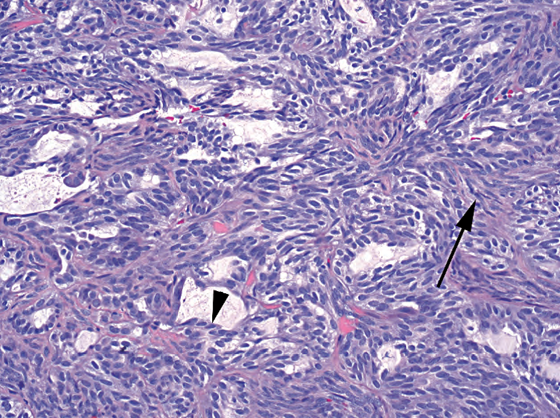
Figure 28.24. Biphasic synovial sarcoma. There are gland-like spaces surrounded by epithelial cells (arrowhead), set in a background of spindle cells (arrow). Monophasic synovial sarcoma lacks the epithelial component and can resemble fibrosarcoma.
图28.24 双相性滑膜肉瘤。有衬覆上皮样细胞的腺样腔隙(箭头),位于梭形细胞(箭)背景中。单相性滑膜肉瘤缺乏上皮样成分,类似纤维肉瘤。
Clear cell sarcoma of soft tissue is one of several translocation tumors linked to the EWS gene. It is called melanoma of soft parts, as it stains with melanoma markers and may even produce melanin. Another EWS tumor is the desmoplastic small round cell tumor, which like it sounds is a small round blue cell tumor in a sclerotic background. The third tumor in this group is Ewing’s sarcoma, which is cytogenetically identical to the peripheral neuroectodermal tumor. It is discussed with bone tumors.
软组织透明细胞肉瘤是与EWS基因相关的几种易位肿瘤之一。又称为软组织黑色素瘤,因为它表达黑色素瘤标记物,甚至可能产生黑色素。另一种EWS肿瘤是促结缔组织增生性小圆细胞肿瘤,听起来像是硬化背景中的小圆蓝细胞肿瘤。第三种肿瘤是Ewing肉瘤,其细胞遗传学与外周神经外胚层肿瘤(PNET)相同,将在骨肿瘤部分讨论。
骨肿瘤(Tumors of Bone)
For tumors of bone, involving bone, or simulating bone, the radiograph is the gross examination. As in vascular lesions, a well-differentiated neoplasm may be classified as benign or low-grade malignant largely by the degree to which it infiltrates or invades surrounding tissue or bone. In bone, this infiltration of the periphery is best assessed by a radiologist. General features are the following:
对于骨肿瘤、累及骨的肿瘤或类似骨的肿瘤,X线检查相当于大体检查。与血管病变一样,分化良好的骨肿瘤可根据其浸润或侵犯周围组织或骨的程度分为良性或低度恶性。骨的周围浸润情况最好由放射科医生评估。一般特点如下:
Benign lesions tend to be clearly defined, well circumscribed, and walled off by a layer of reactive bone (a thin rim on x-ray). Benign entities also tend to evoke a thick and smooth periosteal reaction (thickening).
良性病变往往界限清晰,边界清楚,并由一层反应性骨隔开(X线有一个薄边缘)。良性实体也容易引起厚而光滑的骨膜反应(增厚)。
Aggressive lesions, which include infectious or malignant lesions, tend to be poorly circumscribed, reflecting their infiltration of surrounding bone. Aggressive lesions tend to produce an onion-skin, spiculated, or discontinuous periosteal reaction.
侵袭性病变,包括感染性或恶性病变,往往边界不清,提示对周围骨的浸润。侵袭性病变倾向于产生洋葱皮样、针状或不连续的骨膜反应。
The second major principle is that primary bone tumors are rare and mainly occur in young adults and children. For any patient over 50 years, the first three items in the differential diagnosis for a bony lesion are metastasis, metastasis, and metastasis. Number four is a hematopoietic malignancy such as myeloma.
第二个主要原则是原发性骨肿瘤很少见,主要发生在年轻成人和儿童。对于50岁以上的患者,骨病变鉴别诊断的前三项是转移、转移和转移。第四种是造血系统恶性肿瘤,如骨髓瘤。
成骨肿瘤(Bone-Forming Tumors)
First, how does bone form? In the fetus, the main pathway is by endochondral ossification, in which new bone is laid down in the cartilage scaffolding. However, in the membranous bones of the fetal skull, and in the adult at sites of reparative bone, the first step is the synthesis of osteoid (a salmon-pink acellular matrix) by osteoblasts and its subsequent mineralization with calcium hydroxyapatite. This immature bone has a disorganized collagen framework and is called woven bone. Continuing development and remodeling produce bone with organized sheets of collagen visible as parallel seams within the trabeculae or cortex; this mature configuration is called lamellar bone. Neoplastic or reactive bone is always woven type; fragments of lamellar bone within a lesion must be entrapped native bone.
首先,骨是如何形成的?胎儿主要途径是软骨内骨化,新骨在软骨支架中沉积。胎儿颅骨的膜骨和成人的修复骨部位,第一步是由成骨细胞(或译作骨母细胞)合成类骨质(一种粉红色无细胞基质),随后由羟基磷灰石钙矿化。这种未成熟骨有一个无组织结构的胶原框架,称为编织骨。持续发育和重塑产生骨,骨小梁或皮质内可见有组织结构的成片的胶原平行接合;这种成熟的结构称为板层骨。肿瘤性或反应性骨通常为编织型;病变内的板层骨碎片必须夹住天然骨。
Second, how do we look at bone? Most histologic sections of bone are decalcified, so the pink fragments of “bone” you see are the osteoid left behind. Calcium phosphate itself is dark purple on H&E. In lesions with osteoid formation, which may include anything from reactive metaplastic bone to fibrous dysplasia to osteosarcoma, the osteoid (pink) can be differentiated from collagen or amyloid (also pink) by the process of mineralization, seen as a purple tinge within the seams of osteoid. Dystrophic calcification in soft tissue, such as tumoral calcinosis, is not the same as bone formation. Reactive bone formation, such as in myositis ossificans, is true bone but is not neoplastic.
第二,我们如何观察骨?大多数骨组织切片都脱钙了,所以你看到的“骨”的粉红色碎片就是遗留下来的类骨质。磷酸钙本身在HE上呈深紫色。在类骨质形成的病变中,可能包括从反应性化生骨到纤维发育不良再到骨肉瘤的任何病变,类骨质(粉红色)可以通过矿化过程与胶原或淀粉样(也是粉红色)区分,在骨样接合处可见紫色。软组织中的营养不良性钙化,如肿瘤性钙质沉着症,与骨形成不同。反应性骨形成,如骨化性肌炎,是真正的骨,但不是肿瘤。
The most common benign bone-forming neoplasm is the osteoid osteoma (Table 28.9). This lesion is composed of a small (
最常见的良性成骨肿瘤是骨样骨瘤(表28.9)。该病变是一小块瘤巢(nidus)(
表28.9 成骨肿瘤。
Table 28.9. Bone-forming tumors.
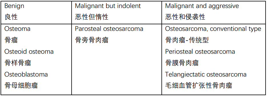
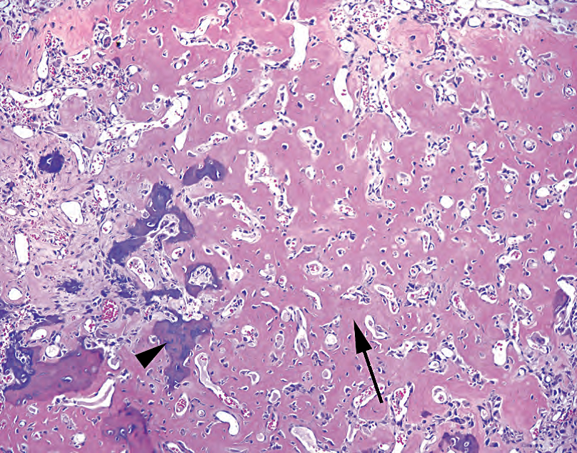
Figure 28.25. Osteoid osteoma or osteoblastoma (depending on size). At the nidus of the lesion, osteoid is laid down in a lace-like pattern (arrow) by benign osteoblasts. The hyaline pink substance can be identified as osteoid by the dark purple seams of mineralization (arrowhead).
图28.25 骨样骨瘤或骨母细胞瘤(取决于大小)。在病变的瘤巢处,良性成骨细胞把类骨质铺成花边样模式(箭)。透明粉红色物质是类骨质,因为有深紫色矿化层(箭头)。
At the other end of the spectrum lies osteosarcoma. The conventional type is a high-grade sarcoma. The usual appearance is that of a high-grade spindle cell neoplasm in which the tumor cells are associated with osteoid deposition (Figure 28.26). The osteoid is laid down in a lace-like pattern, very much like the inside of an osteoma. In fact, a well-differentiated osteosarcoma may be difficult to differentiate from an osteoblastoma. The difference is in the associated population of spindled or atypical cells and in the infiltrative periphery (best appreciated on x-ray). Osteosarcomas may have a wide range of morphologies, including chondroblastic, fibroblastic, and small cell. The unifying and defining feature is the production of osteoid by tumor cells, but osteoid may be sparse and focal. Resections of osteosarcoma are usually done postchemotherapy, at which time the goal is quantifying the amount of viable tumor that remains.
另一端是骨肉瘤。传统型为高级别肉瘤。通常表现为高级别梭形细胞肿瘤,其中肿瘤细胞伴随类骨质沉积(图28.26)。类骨质呈花边状排列,很像骨瘤的内部。事实上,高分化骨肉瘤可能很难与骨母细胞瘤区分。不同之处在于相关的梭形或非典型细胞群和浸润性周围(最好通过X线观察)。骨肉瘤可能有多种形态,包括软骨母细胞型、纤维母细胞型和小细胞型。统一和明确的特征是肿瘤细胞产生类骨质,但类骨质可能稀少和局灶。骨肉瘤的切除通常在化疗后进行,此时检查目标是量化剩余的存活的肿瘤量。
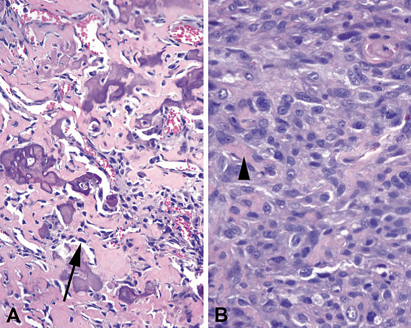
Figure 28.26. Osteosarcoma. (A) The most-well differentiated tumors can be very difficult to distinguish from osteoblastoma by histology alone. The osteoid deposition is similar, except the osteoblasts may appear more hyperchromatic and atypical (arrow). (B) A less differentiated tumor can be difficult to identify as osteosarcoma because of the focal and subtle production of osteoid (arrowhead).
图28.26 骨肉瘤。(A)仅凭组织学很难区分分化最好的骨肉瘤与骨母细胞瘤。类骨质沉积相似,但成骨细胞可能更加深染和更加非典型(箭)。(B)分化较差的骨肉瘤很难识别,因为只有局灶和细微的类骨质(箭头)。
Some variants of osteosarcoma are more indolent. The parosteal osteosarcoma occurs on the surface of the bone, usually behind the knee of a young adult. This tumor is a low-grade sarcoma and is therefore not very cellular or atypical. It may resemble an osteochondroma, with well-formed cartilage and bone, but it is not in continuity with the marrow cavity. The similarly named but quite different periosteal osteosarcoma is also a surface lesion but is primarily chondroblastic and consists of a low-grade spindle cell population with cartilage formation.
骨肉瘤的一些变异型更加惰性。骨旁骨肉瘤发生在骨表面,通常发生在年轻成人的膝盖后面。该肿瘤为低级别肉瘤,因此细胞不太多或非典型不太明显。它可能类似于骨软骨瘤,软骨和骨形成良好,但与骨髓腔不连续。名称相似但完全不同的骨膜骨肉瘤也是一种表面病变,但主要是成软骨细胞,由具有软骨形成的低级别梭形细胞群组成。
成软骨肿瘤(Cartilage-Forming Tumors)
Cartilage-forming tumors produce a characteristic fluffy or concentric-ring pattern of calcification on x-rays (Table 28.10). The osteochondroma is almost diagnosable by x-ray alone, as it stands out from the bone surface like a mushroom. Histologically, it is a bony stalk in continuity with the main marrow space, capped by mature cartilage, looking very much like a duplicated joint surface. Osteochondromas carry a small risk of transformation to chondrosarcoma.
形成软骨的肿瘤在X线上产生特征性蓬松的或同心环状钙化(表28.10)。骨软骨瘤几乎可以只用X线诊断,因为它像蘑菇一样从骨表面突出。组织学上,它是一个与主髓腔连续的骨柄,由成熟软骨覆盖,看起来很像重复的关节面。骨软骨瘤转化为软骨肉瘤的风险很小。
表28.10 成软骨肿瘤。
Table 28.10. Cartilage-forming tumors.

The enchondroma is merely an island of benign, hypocellular, mature cartilage occurring within the marrow space of the bone. It is usually asymptomatic in long bones but is more often found in the small bones of the hands and feet, where it leads to a visible swelling. The tumor consists of mature cartilage, which is a pale blue matrix with varying amounts of calcification (purple) and single chondrocytes sitting in bubble-like lacunae (Figure 28.27).
内生性软骨瘤只是一个良性、少细胞、成熟的软骨岛,发生在骨髓腔内。它通常在长骨中无症状,但在手和脚的小骨中更常见,导致肉眼可见的肿胀。肿瘤由成熟软骨组成,为淡蓝色基质,有不同数量的钙化(紫色),单个软骨细胞位于空泡状腔隙中(图28.27)。
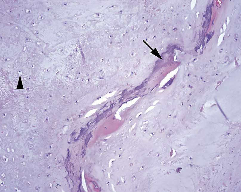
Figure 28.27. Enchondroma. This example shows a sheet of well-differentiated cartilage, with the characteristic blue, somewhat glassy matrix and small chondrocytes embedded in lacunae (arrowhead). The seam of mineralization (arrow) should not be mistaken for osteoid formation. A well-differentiated chondrosarcoma could look very similar histologically.
图28.27 内生性软骨瘤。本例显示一层分化良好的软骨,其特征为蓝色,有点玻璃样基质,小的软骨细胞嵌入陷窝中(箭头)。矿化层(箭)不要误认为是类骨质形成。高分化软骨肉瘤可能很像这种在组织学。
The chondroblastoma is also benign and is notable for a peculiar pattern of calcification that rings the lacunae, creating a chicken-wire or honeycomb effect. The chondromyxoid fibroma is rare but is in the differential diagnosis for a well-differentiated cartilage lesion in bone. It has both a fibrous component and a cartilaginous component.
软骨母细胞瘤也是良性,它有一种特殊的钙化模式。这种钙化模式环绕着陷窝,产生鸡笼样或蜂窝样图像。软骨黏液样纤维瘤罕见,是骨的高分化软骨病变的鉴别诊断之一。它兼有纤维成分和软骨成分。
Chondrosarcoma is typically a mass of atypical but recognizable cartilage, ranging from the low-grade chondrosarcoma (histologically resembling enchondroma) to the high-grade chondrosarcoma (Figure 28.28) based on cellularity, pleomorphism, and mitotic activity. It may be located on the surface of the bone or in the medullary cavity. Features that separate the well-differentiated chondrosarcoma from benign enchondromas include erosion of the inner cortex of bone, entrapment of trabeculae, myxoid change, and a tendency to involve the axial skeleton. Chondrosarcomas are tumors of adults (those in their thirties to fifties).
软骨肉瘤通常是一种非典型软骨病变但可以识别其软骨性质,从低级别软骨肉瘤(组织学类似于内生性软骨瘤)到高级别软骨肉瘤(图28.28)的分级是基于细胞丰富程度、多形性和核分裂活性。它可能位于骨表面或骨髓腔内。区分高分化软骨肉瘤和良性内生性软骨瘤的特征包括侵蚀骨的内层皮质、包裹骨小梁、黏液样改变和往往累及中轴骨。软骨肉瘤是成人肿瘤(30-50岁)。
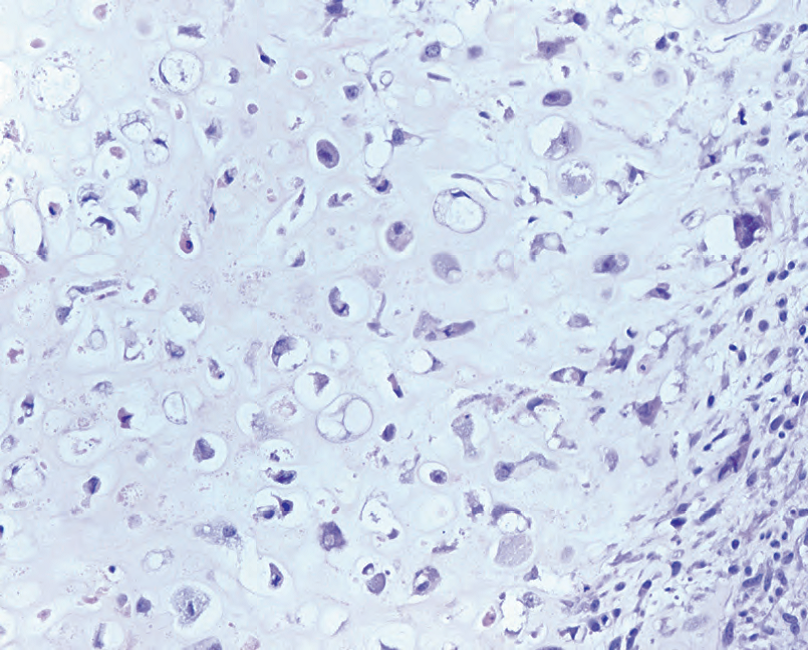
Figure 28.28. Chondrosarcoma. The cartilage matrix resembles normal cartilage, but the chondrocytes are pleomorphic in size and shape.
图28.28 软骨肉瘤。软骨基质与正常软骨相似,但软骨细胞的大小和形状具有多形性。
Chondrosarcomas, even high-grade ones, do not typically have a spindle cell component. A well-differentiated chondrosarcoma with an abrupt transition to high-grade sarcoma (of any pattern) is most likely a dedifferentiated chondrosarcoma. As in the liposarcoma family, this diagnosis relies on seeing the previous or adjacent chondrosarcoma. However, a tumor that shows a gradual transition from cartilaginous areas to high-grade spindle cell areas is more likely a chondroblastic osteosarcoma. Confused? Keep reading.
即使是高级别软骨肉瘤,通常也没有梭形细胞成分。高分化软骨肉瘤突然转变为高级别肉瘤(任何类型)很可能是去分化软骨肉瘤。与脂肪肉瘤家族一样,本诊断依赖于先前或邻近软骨肉瘤的诊断。然而,从软骨区逐渐过渡到高级别梭形细胞区的肿瘤更可能是软骨母细胞型骨肉瘤。困惑吗?请继续。
Osteosarcomas often form cartilage (“chondroblastic”), and chondrosarcomas may mineralize into bone. How do we identify the primary nature of the neoplasm? Chondrosarcoma is a neoplasm of recognizable cartilage without a spindle cell sarcoma component, and the bone formation is through the direct mineralization or ossification of the cartilage, not by osteoid deposition. A cartilage-forming osteosarcoma, on the other hand, should have areas of spindle cells that are producing osteoid. A spindle cell neoplasm intermixed with cartilage, even if the osteoid is not obvious, is more likely to be in the osteosarcoma family.
骨肉瘤通常形成软骨(“软骨母细胞型”),软骨肉瘤可矿化成骨。我们如何确定肿瘤的本质?软骨肉瘤可识别软骨,没有梭形细胞肉瘤成分,骨形成是通过软骨的直接矿化或骨化,而不是类骨质沉积。另一方面,形成软骨的骨肉瘤应该有产生类骨质的梭形细胞区域。梭形细胞肿瘤与软骨混合,即使类骨质不明显,也更有可能属于骨肉瘤家族。
骨的纤维性肿瘤和杂项肿瘤(Fibrous and Miscellaneous Tumors in Bone)
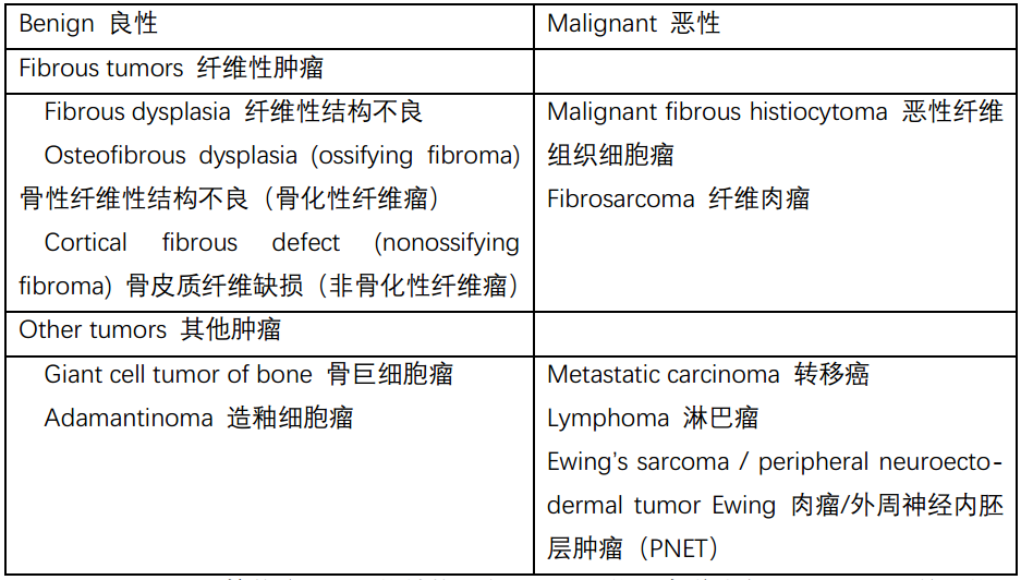
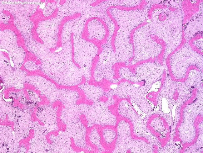
Fibrous dysplasia is a lytic and fibrotic lesion (a developmental abnormality, not really a neoplasm) seen mainly in long bones and craniofacial bones (Table 28.11). M icroscopically, the lesion consists of a low-grade spindle cell population in which thin trabeculae of woven bone are laid down in a distinct pattern resembling (to English speakers) Chinese letters. Unlike typical reactive bone in an inflammatory lesion, osteoblasts are not visible surrounding the trabeculae. Ossifying fibroma is a very similar lesion that occurs in the shins (tibia, fibula) of very young children, only ossifying fibroma does show prominent osteoblastic rimming. Finally, we have the nonossifying fibroma, which is the low-grade fibroblastic population seen in the above lesions, except without the woven bone formation. It is essentially equivalent to a benign fibrous histiocytoma in other sites. The malignant correlates of malignant fibrous histiocytoma (not uncommon) and fibrosarcoma (rare) can occur in bone as well.
纤维结构不良(fibrous dysplasia)是一种溶骨性和纤维性病变(发育异常,并非真正的肿瘤),主要见于长骨和颅面骨(表28.11)。显微镜下,病变由低级别梭形细胞组成,其中编织骨的细小梁以独特的模式排列,对说英语的人来说就像汉字(象形文字)。不同于炎症病变中典型的反应性骨,该病小梁周围未见成骨细胞。骨化性纤维瘤是一种非常类似的病变,发生在小儿胫部(胫骨、腓骨),只有骨化性纤维瘤确实显示明显的成骨细胞边缘。最后,还有非骨化性纤维瘤,这是在上述病变中看到的低级别纤维母细胞群,但没有编织骨形成。实质上相当于其他部位的良性纤维组织细胞瘤。骨也可能发生恶性纤维组织细胞瘤(并不少见)和纤维肉瘤(罕见)。
表28.11 骨的纤维性肿瘤和杂项肿瘤。
Table 28.11. Fibrous and miscellaneous tumors in bone.
(fibrous dysplasia按传统译为纤维结构不良,又称纤维异常增殖症,是不明原因的纤维骨性病变。在骨的瘤样病变中最为常见,约占40%。补充图片如下)
The giant cell tumor of bone is a lytic, destructive lesion seen at the ends of long bones in adults. It is composed of a mixture of osteoclast-like giant cells, often with over 50 nuclei, mixed with a background population of mononuclear cells (the true neoplastic component). Mitoses may be seen, but atypia is not. Giant cells, however, are not a unique feature, as they can be seen in almost any bony lesion. The principal differential is with the giant cell reparative granuloma.
骨巨细胞瘤是一种溶骨性破坏性病变,见于成人长骨末端。它由破骨细胞样巨细胞(通常有50多个核)与背景中的单个核细胞(真正的肿瘤成分)混合组成。可有核分裂,但没有非典型核分裂象。然而,巨细胞并不是独特的特征,因为它们几乎可以在任何骨病变中看到。主要区别在于巨细胞修复性肉芽肿。
Adamantinoma is a rare lesion of the tibia that may be composed of squamous, fibrous, or adamantinomatous (see discussion of craniopharyngioma in Chapter 26) cells. The main reason to know about it is to avoid calling it metastatic carcinoma.
造釉细胞瘤是一种罕见的胫骨病变,可能由鳞状、纤维状或造釉细胞瘤样(见第26章颅咽管瘤的讨论)细胞组成。了解它的主要原因是避免误诊为转移癌。
Ewing’s sarcoma is a tumor of adolescents and young adults and appears as a small round blue cell tumor involving the bone. Like most embryonal-type tumors, the cells have hyperchromatic, round, blue nuclei without prominent nucleoli, high nuclear to cytoplasmic ratios with scant cytoplasm, and prominent necrosis and apoptosis (Figure 28.29). It is classically positive for CD99 and overlaps with the peripheral neuroectodermal tumor, which has identical cytogenetics. The differential diagnosis includes true metastases from other small round blue cell tumors, lymphoma/leukemia, and the small cell variant of osteosarcoma.
尤因肉瘤发生在青少年和年轻成人,表现为累及骨的小圆蓝细胞肿瘤。与大多数胚胎型肿瘤一样,这些细胞的细胞核呈深染、圆形、蓝色,没有明显的核仁,核质比高,细胞质稀少,坏死和凋亡明显(图28.29)。CD99呈典型阳性,与具有相同细胞遗传学的外周神经外胚层肿瘤(PNET)重叠。鉴别诊断包括其他小圆蓝细胞肿瘤、淋巴瘤/白血病和小细胞型骨肉瘤的真正转移。
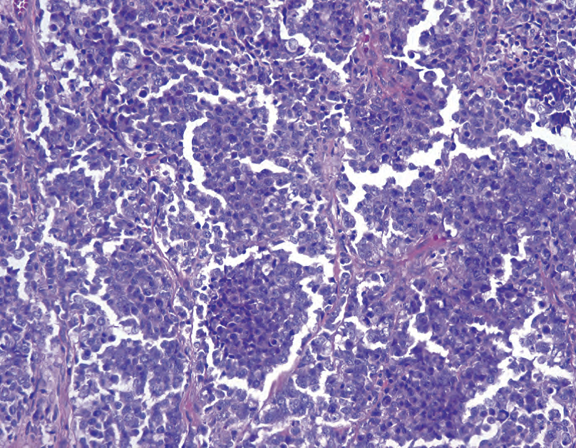
Figure 28.29. Ewing’s sarcoma. The medullary cavity of bone is replaced by a small round blue cell tumor.
图28.29 尤因肉瘤。骨髓腔被小圆形蓝细胞肿瘤所取代。
关节病变(Joint Lesions)
Synovial chondromatosis is one of the few true tumors of the joint space (Table 28.12). It is characterized by the accumulation of nodules of benign cartilage in the synovium.
滑膜软骨瘤病是关节腔的少数真正的肿瘤之一(表28.12)。其特征是滑膜内有良性软骨结节堆积。
The giant cell tumor of tendon sheath actually describes two entities. The diffuse form is also known as pigmented villonodular tenosynovitis, and it is the second of the few lesions to actually involve the joint space. It is composed of a villous or papillary mass of small bland cells, multinucleated giant cells, and foamy macrophages. There are prominent clefted spaces at low power and sometimes pigment (hemosiderin).
腱鞘巨细胞瘤实际上描述了两个实体。弥漫型也称为色素沉着绒毛结节性腱鞘炎,是第二个真正累及关节间隙的少数病变。它由绒毛状或乳头状小细胞团、多核巨细胞和泡沫状巨噬细胞组成。低倍镜下可见明显的裂隙,有时可见色素(含铁血黄素)。
表28.12 关节病变。
Table 28.12. Joint lesions.

The nodular or localized giant cell tumor is also called nodular tenosynovitis, and, aside from presenting as a nodule on a tendon, its appearance is very similar to the diffuse giant cell tumor. The lesion is composed of bland mononuclear cells, giant cells, foamy histiocytes, cleft-like spaces, and hemosiderin, all set in a collagenous stroma. Both of the giant cell tumors of tendon sheath are distinguished from the giant cell tumor of bone by their immunoreactivity to CD68, a histiocyte marker, as well as by their clinical presentation. However, they do resemble each other on H&E.
结节性或局限性巨细胞瘤也称为结节性腱鞘炎,除了表现为肌腱结节外,其形态学很像弥漫性巨细胞瘤。病变由形态温和的单个核细胞、巨细胞、泡沫状组织细胞、裂隙样间隙和含铁血黄素组成,均位于胶原性间质中。这两种腱鞘巨细胞瘤与骨巨细胞瘤的区别在于它们对组织细胞标记物CD68的免疫反应性以及它们的临床表现。然而,他们在HE方面确实很相似。
来源:
The Practice of Surgical Pathology:A Beginner’s Guide to the Diagnostic Process
外科病理学实践:诊断过程的初学者指南
Diana Weedman Molavi, MD, PhD
Sinai Hospital, Baltimore, Maryland
ISBN: 978-0-387-74485-8 e-ISBN: 978-0-387-74486-5
Library of Congress Control Number: 2007932936
© 2008 Springer Science+Business Media, LLC
仅供学习交流,不得用于其他任何途径。如有侵权,请联系删除
本站欢迎原创文章投稿,来稿一经采用稿酬从优,投稿邮箱tougao@ipathology.com.cn
相关阅读
 数据加载中
数据加载中
我要评论

热点导读
-

淋巴瘤诊断中CD30检测那些事(五)
强子 华夏病理2022-06-02 -

【以例学病】肺结节状淋巴组织增生
华夏病理 华夏病理2022-05-31 -

这不是演习-一例穿刺活检的艰难诊断路
强子 华夏病理2022-05-26 -

黏液性血性胸水一例技术处理及诊断经验分享
华夏病理 华夏病理2022-05-25 -

中老年女性,怎么突发喘气困难?低度恶性纤维/肌纤维母细胞性肉瘤一例
华夏病理 华夏病理2022-05-07


共0条评论