外科病理学实践:诊断过程的初学者指南 | 第28章 软组织和骨 上
第28章 软组织和骨(Soft Tissue and Bone)
Tumors of soft tissue are among the most challenging in surgical pathology. There are several reasons for this: they are rare, so you see few in training; they are overlapping in morphology; they do not always obey the principles that help you to identify malignant potential in carcinomas; and each entity has at least three names, four variants, and seven mimickers. However, we will cover some of the names you will hear most commonly. The tumors are broken down into lines of differentiation, with the caveats that there are some tumors that do not differentiate along any known lineage (grouped separately) and that many soft tissue tumors dedifferentiate into the same final common malignant pathway, the entity formerly known as malignant fibrous histiocytoma (MFH). In many instances, MFH is simply a generic dedifferentiated sarcoma, the high-grade form of any one of a number of precursor lesions. The good news is, once it is that high grade, the origin becomes sort of academic.
在外科病理学中,软组织肿瘤具有挑战性。原因:(1)少见,所以你在培训时看得少;(2)形态学重叠;(3)识别癌的恶性潜能的原则,在此不一定适用;(4)每个疾病实体都有多个同义词、变异型和类似病变。我们将介绍一些最常见的肿瘤名字,按分化细胞系进行分类。注意:有些肿瘤的分化细胞系仍然未知(单独分组),许多恶性软组织肿瘤会发生去分化,都会变成最终共同恶性途径,以前称为恶性纤维组织细胞瘤(MFH)。许多情况下,MFH只是去分化肉瘤的一个总称,是由多种前体病变进展而来的一种高级别形式。好消息是,一旦达到如此高的级别,它的起源就变得只是学术性探讨了。
When diagnosing a soft tissue lesion, especially in its initial presentation, you must always walk yourself through the mental game of, “what else could this be?” It is a good habit for any organ system but especially in the field of sarcomas and spindle cell lesions. For lesions that do not look clearly malignant (by which we mean they lack nuclear atypia and necrosis), you must always consider that it might be a reactive lesion. For lesions with bizarre and huge nuclei, despite the malignant look, you must consider benign entities with degenerative atypia (such as an ancient schwannoma). For lesions in or near an organ, such as in visceral sites, you must always ask yourself if it could be a carcinoma masquerading as a sarcoma. For spindle cell lesions anywhere, you must ask yourself if it could be melanoma. Some of these questions require immunostains to answer, some just a skeptical eye.
诊断软组织病变时,尤其是在观察其最初表现时,你必须始终进行“这还能是什么?”的智力游戏,这种诊断思路对任何器官系统都是一个好习惯,尤其是在肉瘤和梭形细胞病变领域。看起来不是明显恶性的病变(意思是缺乏核异型性和坏死),你必须始终认为它可能是反应性损伤。对于具有奇异和巨大核的病变,尽管恶性外观,你必须考虑良性实体与退化非典型性(如退变性神经鞘瘤)。对于器官内或器官附近的病变,如内脏部位,你必须始终扪心自问是否可能是伪装成肉瘤的癌。对于任何地方的梭形细胞病变,你必须问问自己是否可能是黑色素瘤。有些问题需要免疫染色来回答,有些问题只是怀疑的眼光。
(译注:梭形细胞病变:简化的诊断思路
(1)真是恶性吗?
(假的:①无异型,无坏死--良性,反应性?②巨核,怪异核—退变,老化?)
(2)真是恶性。那么,真是肉瘤吗?
(假的:①脏器周围—癌?②任何部位--黑色素瘤?)
(3)真是肉瘤。那么,什么分化?
(免疫组化;即使做了免疫组化也可能没有答案))
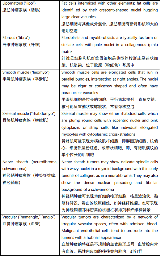
The second question to ask, once you have ordered the cytokeratins and melanoma markers, is “what family of soft tissue does it belong in?” Table 28.1 lists some stereotypical features of different tumor families, seen best in low-grade (well-differentiated) lesions.
一旦你做细胞角蛋白和黑色素瘤标记物的免疫组化,就产生第二个问题:“它属于哪个软组织家族?”表28.1列出了不同肿瘤家族的一些固有特征,在低级别(高分化)病变中最容易看到。
表28.1 不同肿瘤家族的特征。
Table 28.1. Characteristics of tumor families.
高级别肉瘤(High-Grade Sarcomas)
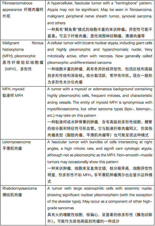
As mentioned, once sarcomas turn high grade, they may take on any number of appearances, regardless of differentiation. Some classic visual patterns are shown in Figure 28.1 and described in Table 28.2.
如前所述,一旦肉瘤变为高级别,无论分化程度如何,都可能出现任意数量上的表现。一些经典的视觉模式如图28.1所示,如表28.2所述。
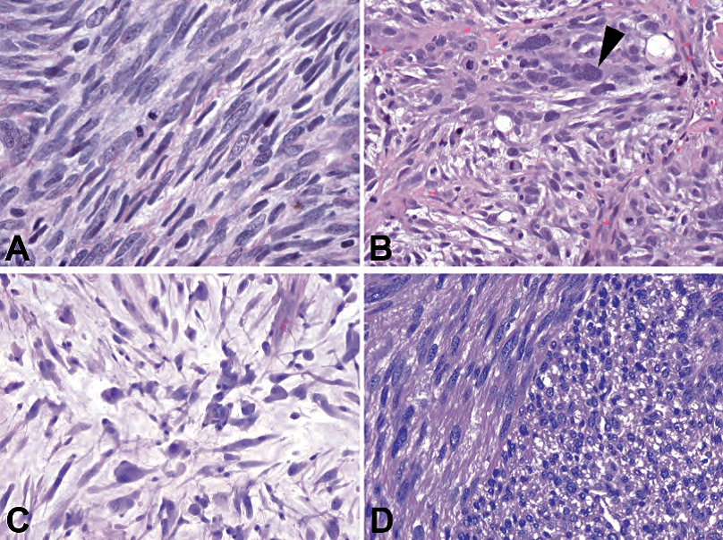
Figure 28.1. High grade sarcomas. (A) Fibrosarcoma, with densely packed hyperchromatic s pindle cells. (B) Pleomorphic MFH, with very large and bizarre cells (arrowhead). (C) Myxoid MFH or myxofibrosarcoma, showing pleomorphic cells in a myxoid background. (D) Leiomyosarcoma, with perpendicular fascicles.
图28.1 高级别肉瘤。(A)纤维肉瘤,有密集的深染的梭形细胞。(B)多形性MFH,有非常大的奇异的细胞(箭头)。(C)黏液样MFH或黏液纤维肉瘤,黏液样背景中的多形性细胞。(D)平滑肌肉瘤,交错成角的束状。
表28.2 高级别肉瘤的特征。
Table 28.2. Features of high-grade sarcoma patterns.
In high-grade sarcomas, the subtype may be clarified through immunostains, history, or the low-grade remnants of the tumor found at the periphery. Identifying the line of differentiation can be helpful in determining prognosis. Solving this puzzle is one of the more interesting games pathologists can play and breaks up the monotony of routine biopsies. However, from a clinical perspective, the oncologist is more concerned about the grade than the type, and, fortunately for all of us, high-grade sarcomas are hard to miss.
高级别肉瘤的亚型,可通过免疫染色、病史或在周围发现的低级别肿瘤残留来阐明。识别分化细胞系有助于判断预后。病理医生有兴趣解决这个难题,活跃了常规活检的氛围。然而,从临床角度来看,肿瘤科医生更关心级别而不是类型,幸运的是,对我们所有人来说,高级别肉瘤很难漏诊。
A reliable clue to a high-grade sarcoma is the presence of malignant nuclei. A sarcoma nucleus has some reproducible features across many tumor types. The nucleus has an irregularly shaped border and has dark, dense, granular chromatin that is fairly evenly distributed throughout the nucleus (Figure 28.2). Unlike carcinoma nuclei, prominent nucleoli and nuclear membranes are not a usual feature. Learning to recognize this sort of atypia is critical in identifying some of the subtle sarcomas.
高级别肉瘤的可靠线索之一是恶性细胞核。肉瘤的细胞核在许多肿瘤类型中具有一些可重复的特征。核外形不规则,有深染、致密、颗粒状染色质,染色质在整个核内均匀分布(图28.2)。与癌细胞核不同,明显的核仁和核膜不是肉瘤的常见特征。学会识别这种异型性对于鉴别某些细微的肉瘤至关重要。
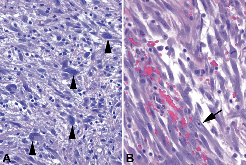
Figure 28.2. The sarcoma cell vs. the reactive cell. (A) Malignant cells in a MFH or other high-grade sarcoma show large nuclei with irregular shapes and very dark chromatin with a coarse texture (arrowheads). It is as though (in fact, it is likely) the nucleus has way too many chromosomes, and the nucleus is swollen and dark with the extra chromatin (truly hyperchromatic). Nucleoli are not usually prominent. (B) Reactive fibroblasts in nodular fasciitis have large nuclei and prominent nucleoli that stand out against a pale nucleus (arrow). The nuclear membrane is smooth and oval.
图28.2 肉瘤细胞与反应性细胞对比。(A)MFH或其他高级别肉瘤中的恶性细胞,大核,形状不规则,染色质非常深染,质地粗糙(箭头)。这就好像(事实上,很可能)细胞核有太多的染色体,而细胞核因额外的染色质(真正的深染)而肿胀和变暗。核仁通常不明显。(B)结节性筋膜炎中的反应性纤维母细胞,有大核和明显核仁,与淡染核形成明显反差(箭头)。核膜光滑,呈椭圆形。
脂肪肿瘤(Tumors of Fat)
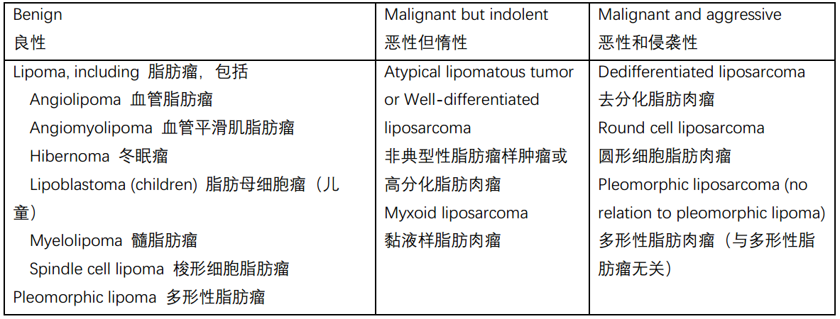
Throughout this chapter, you will find tables listing some of the more common entities, grouped by clinical behavior. Table 28.3 lists some of the common tumors of fat.
本章会列表显示一些较常见的疾病实体,按临床行为分组。表28.3列举一些常见的脂肪肿瘤。
表28.3 常见的脂肪肿瘤。
Table 28.3. Common neoplasms of fat.
The most common soft tissue tumor is the lipoma. A lipoma is defined as a neoplasm of mature fat. It is histologically indistinguishable from ordinary fat; to tell the difference you must know it appeared clinically as a discrete lobular mass. There are many histologic variants of lipoma, classified based on what additional soft tissue component is present, such as the angiolipoma, fibrolipoma, angiomyolipoma, and so forth. The hibernoma is a lipoma of brown fat, in which the fat cells are full of innumerable tiny vacuoles. The lipoblastoma, despite the alarming name, is a benign pediatric tumor of mature fat and benign lipoblasts.
最常见的软组织肿瘤是脂肪瘤。脂肪瘤是指成熟脂肪的肿瘤。组织学上,它与普通脂肪无法区分;真要区分,必须知道它在临床上表现为一个离散的分叶状肿块。脂肪瘤有许多组织学变异型,根据存在的其他软组织成分分类,如血管脂肪瘤、纤维脂肪瘤、血管平滑肌脂肪瘤等。冬眠瘤是棕色脂肪的脂肪瘤,其中脂肪细胞充满无数微小的空泡。脂肪母细胞瘤由成熟脂肪和良性脂肪母细胞组成,为儿科良性肿瘤,尽管其名称令人震惊。
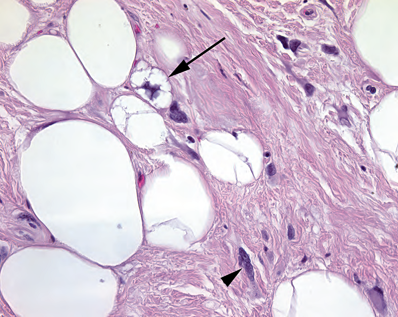
Figure 28.3. Lipoblast. Small fat vacuoles indent the nucleus of this lipoblast (arrow), seen in a welldifferentiated liposarcoma. Other cells within the fibrous septa (arrowhead) have the look of sarcoma cells, with irregular, large, dark nuclei.
图28.3 脂肪母细胞。脂肪母细胞有小的脂肪空泡,挤压细胞核(箭),见于高分化脂肪肉瘤。纤维间隔内的其他细胞(箭头)呈现肉瘤细胞的外观,具有不规则的深染的大核。
There is a lot of fuss about lipoblasts. They are immature fat cells in which the nucleus is star shaped, due to being indented on multiple sides by small bubbles of fat (Figure 28.3). They are often associated with liposarcomas, but they can also appear in the benign lipoblastoma, and they are not necessary for a diagnosis of liposarcoma (more on this later). Note that normal adipocytes are not mitotically active cells, so prominent mitoses are generally seen only in high-grade liposarcomas.
关于脂肪母细胞,可能过分关注了。它是未成熟的脂肪细胞,核呈星形,因为在多个侧面被小的脂肪空泡挤压变形(图28.3)。它们通常与脂肪肉瘤相关,但也可出现在良性脂肪母细胞瘤中,并且对于脂肪肉瘤的诊断不是必需的(稍后详述)。注意,正常脂肪细胞不会核分裂活跃,因此明显的核分裂一般仅见于高级别脂肪肉瘤。
Two types of lipoma are notable for their unusual cytologic features. They are both usually found on the back or neck of elderly men and are noticeably fibrous and nonfatty on low power. These are the pleomorphic lipoma and spindle cell lipoma. The spindle cell lipoma has areas of nondescript spindle cells and collagen and may remind you of a nerve sheath tumor if there is not much fat in the lesion. The pleomorphic lipoma is similar, with the addition of large giant cells and floret cells (wreath-shaped nuclei). These giant cells (Figure 28.4) are an example of a benign lesion simulating malignant atypia; clinical information is helpful in not mistaking these for liposarcomas.
两种脂肪瘤具有不寻常的细胞学特征,即,多形性脂肪瘤和梭形细胞脂肪瘤。它们通常出现在老年男性的背部或颈部,在低倍镜下明显为纤维状和非脂肪状。梭形细胞脂肪瘤有一些难以描述的梭形细胞和胶原区域,如果病变中没有太多脂肪,你可能会想到神经鞘肿瘤。多形性脂肪瘤与之相似,还有大的巨细胞和小花细胞(环状核)。这些巨细胞(图28.4)是典型的假恶性(良性假冒恶性)案例;临床信息有助于避免误认为脂肪肉瘤。
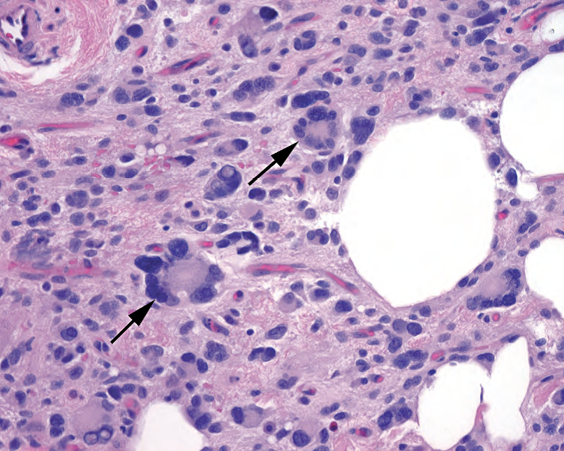
Figure 28.4. Pleomorphic lipoma. This type of benign lipoma is known for having very bizarre stromal cells that mimic sarcoma. The classic cell is the floret cell, with a circular wreath of nuclear lobes (arrows). Their presence suggests the diagnosis of pleomorphic lipoma.
图28.4 多形性脂肪瘤。这种良性脂肪瘤很著名,因为具有非常奇异的貌似肉瘤的间质细胞。典型的细胞是小花样多核巨细胞,多个核叶围成圆环的花环(箭)。其存在提示多形性脂肪瘤的诊断。
The well-differentiated liposarcoma (WDLS) is a tumor of adults. It looks similar to a lipoma on low power except for an increase in fibrous “interstitium” between fat cells and/or fibrous bands (Figure 28.5). A close examination of the fibrous areas reveals hyperchromatic, irregularly shaped nuclei; these are usually large and dark enough to be visible at 4×. Finding a lipoblast is a bonus. A softer feature is an assortment of differently sized fat cells, unlike the monomorphic benign lipoma. The WDLS is so named when it occurs in a nonresectable location, such as the retroperitoneum. By definition, when it occurs on an extremity, it is called an atypical lipoma, as the prognosis in these sites is excellent.
高分化脂肪肉瘤(WDLS)是一种成人肿瘤。低倍镜下,除了脂肪细胞的纤维“间质”增多和/或纤维带增多之外,它看似脂肪瘤(图28.5)。仔细检查纤维区,发现核深染、形状不规则;它们通常大而深染,足以在4倍物镜看到。找到脂肪母细胞是一个额外的收获。较软的特征是各种各样的不同大小的脂肪细胞,不同于形态单一的良性脂肪瘤。发生在不可切除的部位时称为WDLS,如腹膜后。根据定义,发生在四肢时,称为非典型性脂肪瘤,因为这些部位的预后非常好。
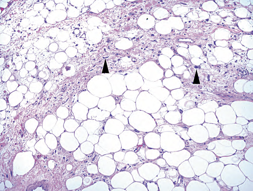
Figure 28.5. Well-differentiated liposarcoma. There is an increased amount of fibrous interstitium between fat cells, and atypical cells stand out at low power (arrowheads).
图28.5 高分化脂肪肉瘤。脂肪细胞之间的纤维间质增多,非典型细胞在低倍镜下明显可见(箭头)。
When the WDLS has been around for a while, especially in a recurrent or occult retroperitoneal lesion, there is a risk of the tumor transforming into a high-grade pleomorphic sarcoma. When this happens, you will see a tumor with well-differentiated lipomatous areas and an abrupt transition to a high-grade tumor (storiform/spindled, MFH-like, or even rhabdo- or leiomyosarcomatous). Regardless of morphology, this is called a dedifferentiated liposarcoma, and the key to diagnosis is recognizing the adjacent WDLS. Because dedifferentiated liposarcoma is the most likely diagnosis in a retroperitoneal pleomorphic sarcoma, if you are grossing such a tumor, be sure to sample anything near the tumor that looks like fat: it may be the well-differentiated component.
WDLS出现一段时间后,尤其是在复发性或隐匿性腹膜后病变中,肿瘤有转化为高级别多形性肉瘤的风险。这种情况发生时,你会看到肿瘤有一个高分化的脂肪瘤样区域,突然转变为高级别肿瘤(席纹状/梭形、MFH样,甚至横纹肌肉瘤样或平滑肌肉瘤样)。不管形态学如何,都称为去分化脂肪肉瘤,诊断的关键是识别相邻的WDLS。因为去分化脂肪肉瘤是腹膜后多形性肉瘤中最有可能的诊断,如果你正在大体检查,在肿瘤附近看似脂肪的东西一定要取材:它可能是高分化成分。
Myxoid liposarcoma is a different type of low-grade liposarcoma. It is not as clearly fatty as the WDLS, and the low-power impression is that of a gelatinous tumor with scattered fat cells and a stereotypical capillary network that has been compared to chicken-wire (Figure 28.6). These vessels are very delicate, and, unlike normal capillaries, they have little substance to their walls; they appear as a naked sleeve of endothelium stretched through the tumor. The tumor cells themselves are small spindled or rounded cells and lipoblasts, without the large atypical cells of the WDLS.
黏液样脂肪肉瘤是另一种的低级别脂肪肉瘤。它不像WDLS那样有明显脂肪,低倍镜下的印象是黏液样肿瘤,有散在的脂肪细胞和特征性毛细血管网,后者像鸡笼(图28.6)。这些血管非常纤细,与正常的毛细血管不同,它们的血管壁几乎没有物质;就像裸露的内皮细胞袖套,穿过肿瘤。肿瘤细胞本身是小的梭形或圆形细胞和脂肪母细胞,没有WDLS那样的非典型大细胞。
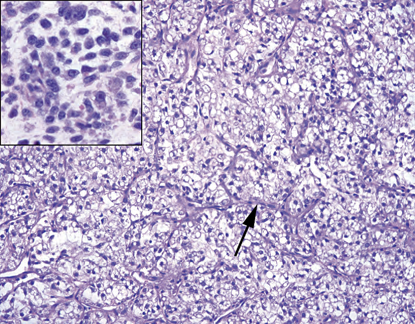
Figure 28.6. Myxoid liposarcoma. The fatty component may be very subtle in myxoid liposarcoma; the vessels are more often the tip off. The vasculature is composed of a delicate network of very thin capillaries with three- and four-way branch points, similar to chicken-wire (arrow). The cell population is composed of small cells, which may have fat vacuoles in them, and a myxoid background. Large atypical cells should not be present. Inset: Areas of closely packed small cells are indicative of round cell differentiation.
图28.6 黏液样脂肪肉瘤。脂肪成分可能非常细微;血管通常是诊断线索。血管由非常细的毛细血管组成,并形成精致的网。毛细血管具有三向和四向分支点,就像鸡笼(箭)。细胞群由小细胞组成,可能有脂肪空泡,位于黏液样背景中。不应出现非典型大细胞。插图:紧密排列的小细胞区域提示圆形细胞分化。
The myxoid liposarcoma can also transform into a higher grade lesion. When the small uniform cells become very densely packed and obscure the vascular pattern, it is indicative of round cell differentiation. The presence of more than 5% round cell differentiation is worth noting; a tumor with more than 75% is called a round cell liposarcoma.
黏液样脂肪肉瘤也可转化为更高级别病变。当小而均匀的细胞变得非常密集,使血管模式变得模糊时,表明细胞分化为圆形。圆形细胞分化超过5%没有意义;超过75%的肿瘤称为圆形细胞脂肪肉瘤。
The rare pleomorphic liposarcoma describes a high-grade tumor with extremely bizarre pleomorphic lipoblasts. It differs from the dedifferentiated liposarcoma in that it is still recognizable as a lipomatous tumor. It does not arise from, or have any relation to, the pleomorphic lipoma.
多形性脂肪肉瘤是一种罕见的高级别肿瘤,具有极其奇异的多形性脂肪母细胞。它不同于去分化脂肪肉瘤,它仍然可以识别为脂肪性肿瘤。它不是由多形性脂肪瘤引起的,也与多形性脂肪瘤无关。
纤维肿瘤和黏液样肿瘤(Fibrous Tumors and Myxoid Tumors)
The fibroblast and the myofibroblast are ubiquitous cells, in charge of the reparative changes that take place in every part of the body. In resting state, they are fusiform to stellate cells with oblong pale nuclei, and they lay down a collagen matrix. Their job is to proliferate, and therefore mitotic activity is not unusual in reactive lesions. Although myofibroblasts may stain for actin (and are occasionally mistaken for smooth muscle), in general immunostains are not helpful in this tumor family.
纤维母细胞和肌纤维母细胞是普遍存在的细胞,负责身体各个部位发生的修复性变化。在静息状态下,它们呈梭形至星芒状,核呈淡染的长方形。它们产生胶原基质。它们的工作是增殖,因此核分裂活性在反应性病变中并不罕见。虽然肌纤维母细胞可以染actin(偶尔被误认为平滑肌),但一般来说,免疫染色对该肿瘤家族没有帮助。
反应性病变(Reactive lesions)
Before we discuss the true neoplasms, we will review the collection of reactive (inflammatory or posttraumatic) lesions that present as tumors (lumps). Keloid is a common fibrous lesion, occurring at a site of trauma. It is similar to the normal fibroblastic proliferation of a dermal scar, except for its large size and characteristic thick cords of collagen called keloidal collagen. It is clinically recognizable and not usually a diagnostic dilemma.
在讨论真正的肿瘤之前,我们先回顾一组表现为肿瘤(肿块)形式的反应性(炎症或创伤后)病变。瘢痕疙瘩是一种常见的纤维病变,发生在创伤部位。它类似于真皮瘢痕的正常纤维母细胞增殖,除了它的尺寸大和特征性厚胶原索(称为瘢痕疙瘩胶原)。它在临床上可以识别,通常不是诊断难题。
Nodular fasciitis, on the other hand, may simulate a neoplasm clinically and is therefore more challenging for the pathologist. It is classically a rapidly growing lesion, sometimes associated with known trauma, sometimes not. On low power, it is a fairly circumscribed lesion with a hypercellular periphery, and it has a heterogeneous, as opposed to monomorphic, look. A microcystic appearance is classic. On high power the fibroblasts show a “tissue culture” appearance (fusiform to stellate with distinct cytoplasmic processes), and they float in a myxoid background with surrounding red blood cells and lymphocytes (Figure 28.7). Older lesions may become more dense, collagenized, and pink and may resemble a fibromatosis (see later) with chronic inflammation. There should be no nuclear atypia, but you will see mitotic activity.
另一方面,结节性筋膜炎在临床上可能假冒肿瘤,因此对病理医生来说更具挑战性。它是典型的快速增长的病变,有时与已知的创伤有关,有时不相关。在低倍镜下,它具有相当清楚边界,外周细胞丰富,并且它具有异质性,而不是单一形态的外观。微囊表现是典型的。在高倍镜下,纤维母细胞呈现“组织培养”外观(梭形到星芒状,具有清楚的细胞质突起),它们漂浮在黏液样背景中,周围有红细胞和淋巴细胞(图28.7)。较老的病变可能变得更致密、胶原化和粉红色,可能类似于伴有慢性炎症的纤维瘤病(见下文)。应该没有核异型性,但你会看到核分裂活性。
The biggest pitfall in nodular fasciitis is misinterpreting the patchy high cellularity and mitotic activity for a sarcoma. The clinical history is helpful, as is recognizing the reactive versus malignant nuclear features (something that takes practice).
结节性筋膜炎的最大诊断陷阱,就是把肉瘤的斑片状高度富于细胞的和核分裂活跃的区域误认为肉瘤。临床病史很有帮助,认识反应性与恶性核特征的区别也有助于诊断(需要实践经验)。
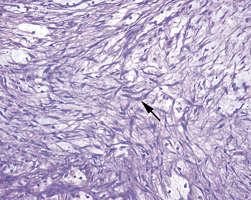
Figure 28.7. Nodular fasciitis. In this field the inflammatory component is not prominent, but the “tissue culture” pattern is seen clearly, with fusiform and stellate fibroblasts stretching delicate processes through the myxoid background (arrow).
图28.7 结节性筋膜炎。在这个视野,炎症成分不明显,但“组织培养”模式清晰可见,梭形和星芒状纤维母细胞在黏液样背景中伸展出精细的突起(箭)。
Proliferative fasciitis is similar to nodular fasciitis except with the addition of large pink ganglionlike cells. Proliferative myositis is essentially the same lesion but in an intramuscular location.
增生性筋膜炎与结节性筋膜炎相似,只是还有大的粉红色神经节样细胞。增生性肌炎本质上是相同的病变,但位于肌肉内。
Myositis ossificans is a variant of proliferative myositis that shows reactive bone formation.
骨化性肌炎是增生性肌炎的一种变异型,表现为反应性骨形成。
Inflammatory myofibroblastic tumor has gone by many names (inflammatory pseudotumor, inflammatory fibrosarcoma, plasma cell granuloma, others), but in this chapter it will be shortened to IMT. While long considered a reactive lesion, occasional cases have spread aggressively and even metastasized. Therefore, it is beginning to be regarded as a neoplasm, and will be included below, despite its histologic similarity to nodular fasciitis.
炎性肌纤维母细胞瘤有许多名称(炎性假瘤、炎性纤维肉瘤、浆细胞肉芽肿等),但在本章中简称为IMT。虽然长期以来认为它是反应性病变,但偶尔也会发生侵袭性扩散甚至转移。因此,尽管其组织学与结节性筋膜炎相似,但它已开始被视为肿瘤,并将在下文中予以介绍。
肿瘤(Neoplasms)
The prototypical benign fibroblastic lesion is the fibromatosis (Table 28.4). This is a bland and indistinct tumor composed of normal-looking fibroblasts: fascicles of pink cells with pale tapering nuclei in a collagenous background (Figure 28.8). The very pale nuclei make the capillaries stand out and appear dark in comparison. It is very infiltrative around the edges, much like a normal scar. Superficial fibromatoses can occur on the palm (palmar fibromatosis, Dupuytren’s contracture), sole (plantar, Ledderhose disease), or penis (Peyronie’s disease), where they are benign but can recur. Axial or deep fibromatoses, such as on the chest wall or mesentery, are typically more aggressive in their expansion and are called desmoid tumors. The desmoid tumors are characterized by a specific immunohistochemical trait, the accumulation of β-catenin in nuclei.
良性纤维母细胞病变的原型是纤维瘤病(表28.4)。这是一种形态学温和、模糊不清的肿瘤,由看起来正常的纤维母细胞组成:粉红色细胞束,淡染的锥形核,位于胶原背景中(图28.8)。核非常淡染,使得毛细血管明显和深染。边缘呈明显的侵袭性,很像正常的疤痕。浅表性纤维瘤病可发生在手掌(掌纤维瘤病、Dupuytren挛缩)、脚底(跖纤维瘤病、Ledderhose病)或阴茎(Peyronie病)等部位,这些都是良性但可能复发(译注:2020年WHO分类中ICD-O编码8813/1)。躯干或深部纤维瘤,如胸壁或肠系膜上的纤维瘤,通常在扩散时更具侵袭性,称为硬纤维瘤。硬纤维瘤具有特殊的免疫组化表达,即β-catenin在细胞核中的积累。
表28.4 纤维肿瘤和粘液样肿瘤。
Table 28.4. Fibrous and myxoid neoplasms.
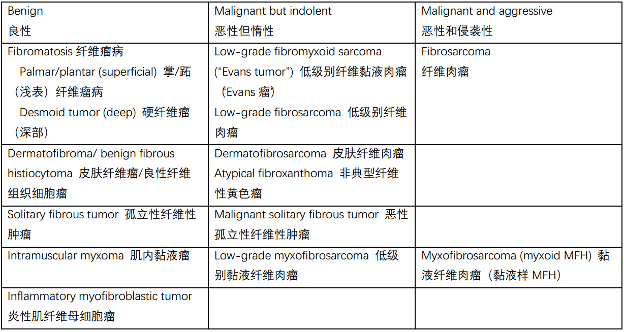
Low-grade fibromyxoid sarcoma is one of those most troublesome entities; it simulates a benign lesion (fibromatosis) yet has metastatic potential. It may appear hypocellular, myxoid, or vaguely storiform, much like a fibromatosis.
低级别纤维黏液样肉瘤是最麻烦的实体之一;它貌似良性病变(纤维瘤病),但具有转移潜能。它可能表现为细胞少、黏液样或隐约的席纹状,很像纤维瘤病。
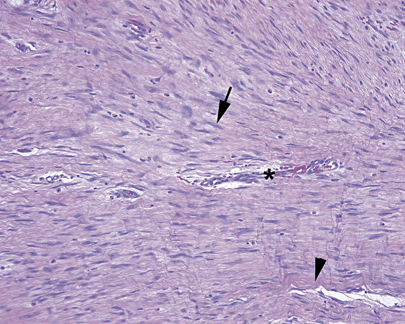
Figure 28.8. Fibromatosis. The cells in this lesion are pale and indistinct, with the small wavy nuclei (arrow) noticeably hypochromatic relative to the endothelial cells of the nearby capillary (a good internal control; asterisk). There is abundant collagen in the stroma (arrowhead).
图28.8 纤维瘤病。病变中的细胞淡染、不清楚,与附近毛细血管(良好的内部控制;星号)的内皮细胞相比,小的波纹状核(箭)明显淡染。基质中有丰富的胶原(箭头)。
Fibrosarcoma is the high-grade endpoint of this spectrum of lesions. It is the classic pure “herringbone” lesion, with fascicles alternating in a zigzag pattern. There is no significant collagen or inflammation to speak of. It has a high mitotic rate, but the cells are not particularly atypical: the nuclei tend to be monomorphic, oval, and euchromatic (Figure 28.9). It is mainly the cellular density and mitotic activity that set this lesion apart as malignant.
纤维肉瘤是这个病变谱系中的高级别终点。这是典型的纯“鲱鱼骨”病变,细胞束以之字形交替排列。没有明显的胶原或炎症。核分裂率高,但细胞异型性并不特别明显:核往往形态单一、椭圆形和正常染色(图28.9)。主要根据细胞密集和核分裂活性判定为恶性。
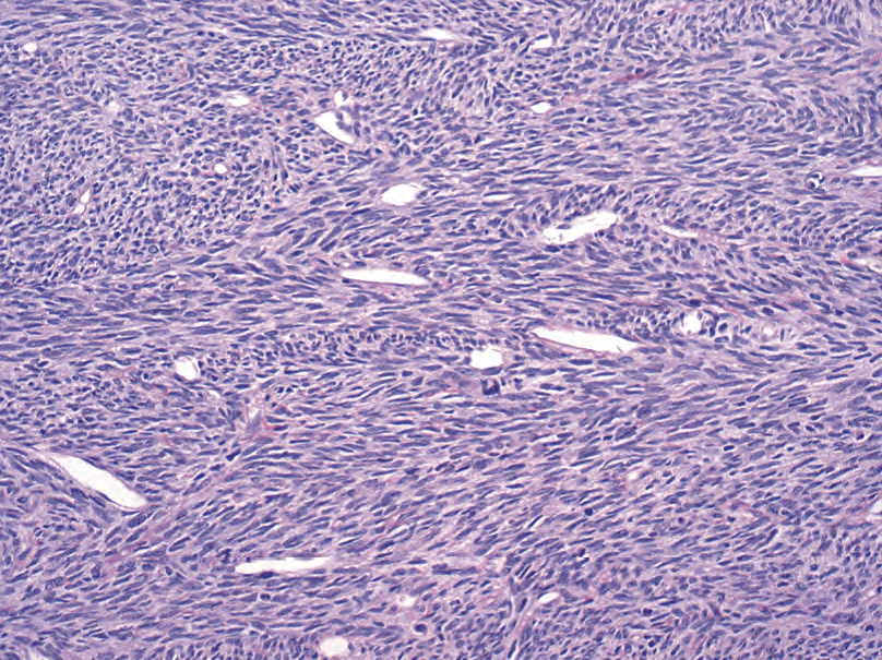
Figure 28.9. Fibrosarcoma. This field shows the typical herringbone pattern of fibrosarcoma, with zigzagging bands of spindle cells. Many other tumors can have this pattern.
图28.9 纤维肉瘤。这个视野显示纤维肉瘤典型的鲱鱼骨模式,梭形细胞呈之字形带状排列。许多其他肿瘤也有这种模式。
However, what looks like fibrosarcoma is not usually fibrosarcoma. True fibrosarcoma is quite rare, while its imitators, especially malignant peripheral nerve sheath tumor and synovial sarcoma, are more common. Therefore, fibrosarcoma is a diagnosis of exclusion.
然而,看似纤维肉瘤的,通常不是纤维肉瘤。真正的纤维肉瘤非常罕见,其假冒病变更为常见,尤其是恶性周围神经鞘瘤和滑膜肉瘤。因此,纤维肉瘤是一种诊断排除。
The solitary fibrous tumor is included here because of its resemblance to fibroblastic tumors, but in truth the type of differentiation is not known. The solitary fibrous tumor has a unique staining pattern (CD34, CD99, and bcl-2) and typically arises from serosal surfaces. Because of its association with the pleura, it was once called the “benign mesothelioma.” On low power, the tumor is described as having a patternless pattern, which evidently means nonstoriform-nonherringbone-nonfascicular. The swirling mass of uniform cells is reminiscent of ovarian stroma, but appears more pink due to abundant collagen (Figure 28.10). Collagen is laid down in parallel bundles, and the cellularity varies from one field to the next. The vessels are of the “staghorn” type, meaning they are gaping, branching vessels without an appreciable wall thickness: the tumor appears to extend right up to the endothelium. A mitotic rate of more than 4 per 10 high-power fields (hpf) suggests a malignant solitary fibrous tumor.
由于与纤维母细胞瘤相似,在此讨论孤立性纤维性肿瘤(SFT),但实际上分化类型尚不清楚。SFT具有独特的染色模式(CD34、CD99和bcl-2),通常发生在浆膜表面。由于它与胸膜有关,曾被称为“良性间皮瘤”。在低倍镜下,肿瘤被描述为“无模式的模式",这显然意味着非席纹状非鲱鱼骨非束状结构。形态均匀的细胞形成漩涡状细胞团,就像卵巢间质,但由于富含胶原而显得更红(图28.10)。胶原以平行束状排列,各个视野的细胞密度不相同。血管为“鹿角”型,即,开放的分支血管,没有明显的厚壁血管:肿瘤似乎一直延伸到内皮细胞。核分裂率超过4/10HPF提示为恶性SFT。
(译注:2020年WHO分类中,SFT归入纤维母细胞肿瘤,临床行为有良性、中间性和恶性,编码分别为8815/0、8815/1和8815/3)
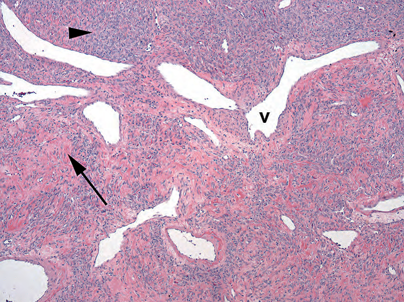
Figure 28.10. Solitary fibrous tumor. The most noticeable features at low power are the staghorn vessels (v), which this tumor shares with the related hemangiopericytoma. The tumor is composed of areas of small nondescript spindle cells (arrowhead) and collagenous stroma (arrow). The pattern of the spindle cells is described as “patternless,” meaning somewhat chaotic.
图28.10 孤立性纤维性肿瘤。低倍镜下最显著的特征是鹿角状血管(v),这与相关的血管外皮细胞瘤相同。肿瘤由无法描述的小梭形细胞区域(箭头)和胶原基质区域(箭)组成。梭形细胞的模式被描述为“无模式”,这意味着有些混乱。
皮肤的纤维性肿瘤(Fibrous Tumors of the Skin)
The benign fibrous/fibrohistiocytic tumor of the dermis is the dermatofibroma or benign fibrous histiocytoma. It appears clinically as a little nodule, and at low power you see a diffuse, hazy, indistinct “blueness” occupying and expanding the dermis. On higher power, the dermatofibroma shows a textbook storiform pattern, with spindly cells arranged in little radial sunbursts, like spokes on a wheel (see Figure 27.23 in Chapter 27). There is also usually accompanying inflammation, including lymphocytes, plasma cells, and foamy macrophages.
真皮的良性纤维/纤维组织细胞肿瘤是皮肤纤维瘤或良性纤维组织细胞瘤。临床上表现为小结节,低倍镜下可见弥漫、云雾状、模糊的“蓝色”占据并扩张真皮。在高倍镜下,皮肤纤维瘤呈教科书式的席纹状模式,梭形细胞排列成小的放射状,就像车轮上的辐条(参见第27章图27.23)。通常伴有炎症,包括淋巴细胞、浆细胞和泡沫状巨噬细胞。
The malignant, albeit indolent, counterpart is the dermatofibrosarcoma protuberans (DFSP). This lesion is also distinctly storiform, and the tumor cells, as in the dermatofibroma, are monomorphic, fusiform, and just slightly hyperchromatic. However, the DFSP penetrates more deeply and classically infiltrates the subcutaneous fat, wrapping around fat cells in a distinctive pattern (see Chapter 27). In contrast to the dermatofibroma, the DFSP is ironically more uniform in cytology and lacks the associated inflammatory cells.
恶性但惰性的对应物是隆突性皮肤纤维肉瘤(DFSP)。这种病变也是明显的席纹状,肿瘤细胞正如皮肤纤维瘤,是单形的梭形细胞,只是稍微深染。然而,DFSP浸润更深,通常浸润到皮下脂肪,以独特的模式包裹脂肪细胞(见第27章)。与皮肤纤维瘤相比,矛盾的是DFSP在细胞学上更为一致,并且缺少相关的炎性细胞。
The skin also has its own MFH variant, called an atypical fibroxanthoma. Despite being histologically equivalent to a pleomorphic MFH, this superficial tumor is easily resected and therefore has a good prognosis.
皮肤也有自己的MFH变异型,称为非典型纤维黄色瘤。尽管组织学相当于多形性MFH,但这种浅表肿瘤很容易切除,因此预后良好。
黏液样肿瘤(Myxoid Tumors)
The myxoid lesions included here are those that are not myxoid variants of other tumor types (such as myxoid liposarcoma). Many different lesions may converge on the myxoid phenotype, however. What we call myxoid is really the accumulation of hyaluronic acid, a gel-like substance that is essentially a form of solid water in the body (as seen in tissue edema). It may appear clear to a very pale blue on routine stains. A myxoid differential diagnosis would include myxoma, angiomyxoma, neurofibroma, and nodular fasciitis (all benign) and myxoid MFH (myxofibrosarcoma), myxoid liposarcoma, myxoid chondrosarcoma, myxoid leiomyosarcoma, and the low-grade fibromyxo- and myxofibro- entities (all malignant). You would also need to exclude tumors that may appear myxoid but are not, including chordoma, cartilaginous tumors, and epithelial mucinous tumors.
这里讨论的黏液样病变不是其他肿瘤类型的黏液样变异型(如黏液样脂肪肉瘤)。然而,许多不同的病变可呈黏液样表型。所谓的黏液样物质实际上是透明质酸的积累,一种凝胶状物质,本质上是身体内固体水的一种形式(如组织水肿)。常规染色,它可能呈现非常淡的蓝色。黏液样肿瘤的鉴别诊断包括黏液瘤、血管黏液瘤、神经纤维瘤、结节性筋膜炎(均为良性)和黏液样MFH(黏液纤维肉瘤)、黏液样脂肪肉瘤、黏液样软骨肉瘤、黏液样平滑肌肉瘤,以及低级别黏液纤维肉瘤和黏液纤维肉瘤。你还需要排除可能呈黏液样,但并非黏液样肿瘤,包括脊索瘤、软骨性肿瘤和上皮性黏液肿瘤。
The intramuscular myxoma is a benign and nearly acellular lesion that presents as a gelatinous mass within a muscle, usually in women. There are rare small stellate cells without atypia. What separates the benign myxoma from other more worrisome lesions is its virtual lack of capillaries.
肌内黏液瘤是一种良性的几乎无细胞的病变,通常表现为女性患者肌肉内的凝胶状肿块。罕见的小星芒状细胞没有异型性。良性黏液瘤与其他更令人担忧的病变的区别在于它实际上缺乏毛细血管。
Myxofibrosarcoma is a high-grade tumor also known as myxoid MFH, and its low-grade counterpart is the low-grade myxofibrosarcoma (not to be confused with the fibromatosis-like low-grade fibromyxoid sarcoma!). The myxofibrosarcomas are tumors that are prominently myxoid but that have an increasing cellularity, nuclear pleomorphism, and mitotic rate compared to myxoma. Because of prominent vessels and bubbly cells (pseudolipoblasts), they may be mistaken for myxoid liposarcoma. However, the vessels are different. Myxofibrosarcoma vessels are “curvilinear,” which means they make short thick arcs in the tumor, and the tumor cells appear to drip off of them like wax from a candle (Figure 28.11). Compare these to the delicate branching capillaries of the myxoid liposarcoma (see Figure 28.6). The nuclei of myxofibrosarcoma also set them apart; they are hyperchromatic and pleomorphic, unlike the uniform nuclei of the myxoid liposarcoma.
黏液纤维肉瘤是一种高级别肿瘤,也称为黏液样MFH,其低级别对应物是低级别黏液纤维肉瘤(不要与纤维瘤病样低级别纤维黏液样肉瘤混淆!)。黏液纤维肉瘤是一种明显的黏液样肿瘤,但与黏液瘤相比,其细胞数量、核多形性和核分裂率增加。由于明显的血管和空泡状细胞(假脂母细胞),它们可能误认为是黏液样脂肪肉瘤。然而,血管是不同的。黏液纤维肉瘤血管呈“曲线”,这意味着它们在肿瘤中形成短而粗的弧形,肿瘤细胞似乎像蜡烛上的蜡一样从血管中滴下(图28.11)。对比黏液样脂肪肉瘤的精致的分支状毛细血管(见图28.6)。黏液纤维肉瘤的细胞核呈深染和多形性,也能鉴别黏液样脂肪肉瘤的均匀细胞核。
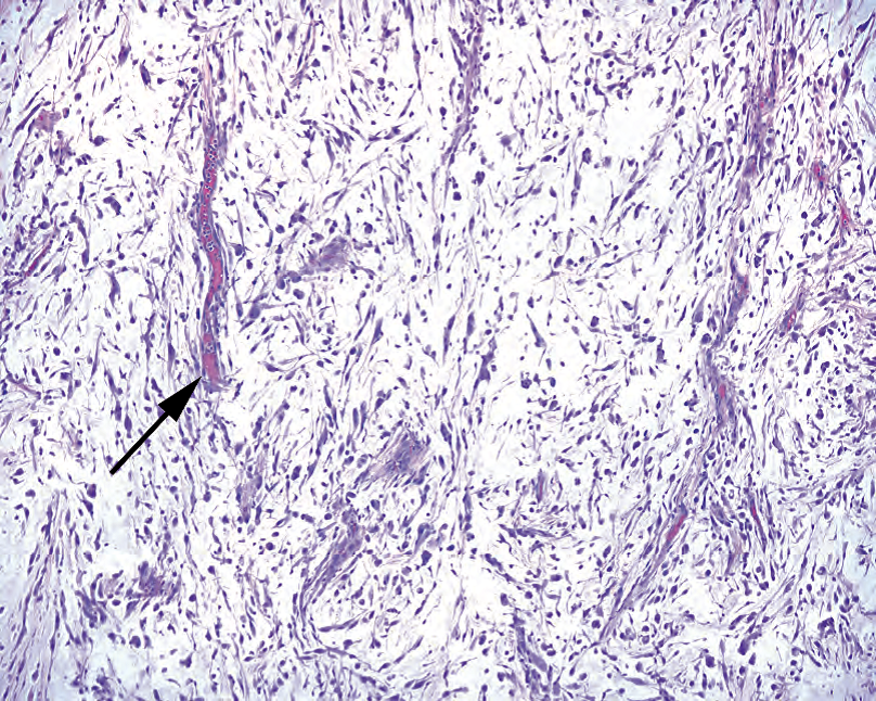
Figure 28.11. Myxofibrosarcoma. Although the cells here resemble those of pleomorphic malignant fibrous histiocytoma, the stroma is myxoid, and the vessels are unique (arrow). They appear as short arcs or segments, unlike the complex branching vessels of myxoid liposarcoma, and the tumor cells are intimately associated with the vessels, like wax dripping down the side of a candle.
图28.11 黏液纤维肉瘤。虽然这里的细胞类似于多形性MFH,但基质是黏液样的,血管是独特的(箭)。与黏液样脂肪肉瘤的复杂分支血管不同,它们呈短弧形或节段,肿瘤细胞与血管密切相关,就像蜡从蜡烛边滴下一样。
Inflammatory myofibroblastic tumor (IMT) is a neoplasm of mainly young people, often arising in the abdominal cavity. It is a proliferation of plump fibroblasts with abundant associated inflammation, especially plasma cells. It is very similar in appearance to a nodular fasciitis in that there are tissue culture–like fibroblasts in a myxoid, granulation tissue–like background (Figure 28.12). It differs by its visceral location and prominence of plasma cells (not seen in nodular fasciitis). The hypercellularity may be very worrisome for a high-grade sarcoma. However, while the nuclei may be enlarged, with prominent nuclear membranes or large nucleoli, you should not see the irregularly shaped hyperchromatic nuclei of MFH. Many cases show immunoreactivity for ALK.
炎性肌纤维母细胞瘤(IMT)是一种主要发生于年轻人的肿瘤,常发生于腹腔。它是一种丰满的纤维母细胞增殖,伴有大量相关炎症,尤其是浆细胞。它看起来很像结节性筋膜炎:组织培养样纤维母细胞,位于黏液样、肉芽组织样背景中(图28.12)。其不同之处在于内脏位置和浆细胞的明显(结节性筋膜炎中未见)。细胞丰富可能令人担心高级别肉瘤来。然而,尽管核可能增大,有明显核膜或大核仁,但不应看到MFH那种形状不规则的深染核。许多病例对ALK有免疫反应。
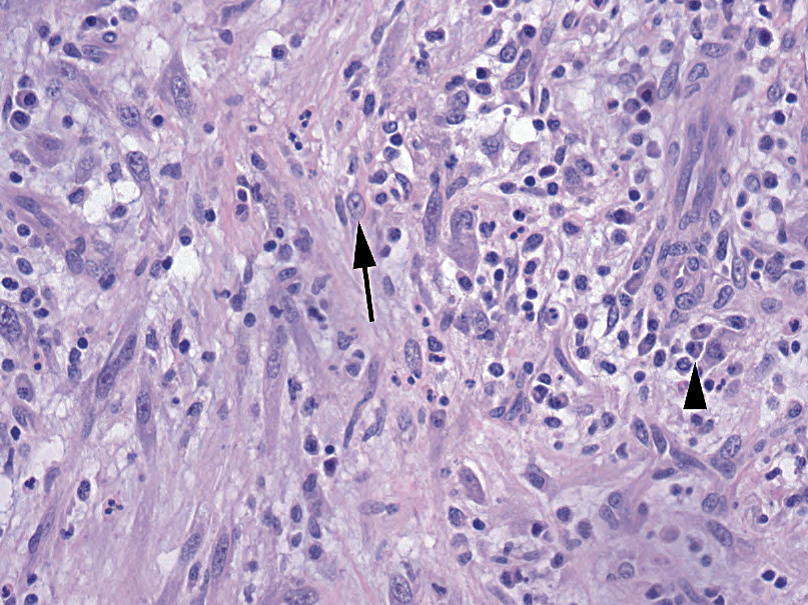
Figure 28.12. Inflammatory myofibroblastic tumor. The tumor is composed of a network of reactivelooking fibroblasts (arrow), capillaries, and inflammation, especially plasma cells (arrowhead).
图28.12 炎性肌纤维母细胞瘤。肿瘤由看似反应性的纤维母细胞(箭)、毛细血管和炎症,尤其是浆细胞(箭头)组成。
平滑肌肿瘤(Smooth Muscle Tumors )
There are no reactive smooth muscle lesions, so we will go straight to neoplasms (Table 28.5). Smooth muscle neoplasms may be positively identified by immunoreactivity to actin and desmin but may sometimes show spurious cytokeratin or EMA staining.
没有反应性平滑肌病变,因此我们将直接讨论肿瘤(表28.5)。平滑肌肿瘤可通过actin和desmin的免疫反应阳性来识别,但有时可显示细胞角蛋白或EMA染色的假阳性。
表28.5 平滑肌肿瘤。
Table 28.5. Smooth muscle neoplasms.

The leiomyoma should be familiar, as it is identical to the uterine tumor. It can occur as a primary neoplasm in cutaneous, gastrointestinal, and other sites. However, unlike in the uterus, in these body sites there is a very low threshold for bumping the lesion up to leiomyosarcoma. In general, greater than 1 mitosis per 10 hpf is worrisome.
软组织平滑肌瘤应该很熟悉,因为它与子宫平滑肌瘤相同。它可以发生于皮肤、胃肠道和其他部位。然而,与子宫不同,这些部位诊断为平滑肌肉瘤的阈值非常低。总的来说,核分裂象超过1/10HPF是令人担忧的。
The leiomyoma is characterized by long parallel bundles of smooth muscle cells that intersect at right angles, such that some are seen longitudinally and some cut in cross section. The nuclei are often described as cigar or box-car shaped, with blunt ends. You may also see c orkscrew nuclei, which appear twisted about themselves and are associated with the contracted state. Paranuclear vacuoles are common (Figure 28.13).
平滑肌瘤的特征是长的平行的平滑肌细胞束,以直角相交,有的纵向可见,有的横切面可见。细胞核通常被描述为雪茄或车厢形,末端钝圆。你也可以看到螺旋核,它看起来是扭曲的,与收缩状态有关。核旁空泡常见(图28.13)。
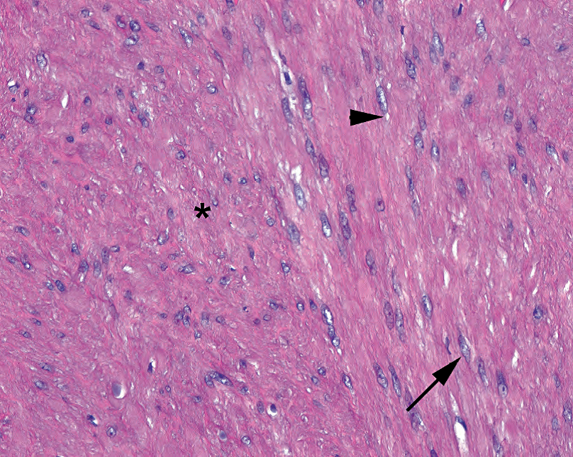
Figure 28.13. Leiomyoma of colon. As in the leiomyoma of the uterus, there are smooth muscle cells in bundles running parallel to and perpendicular to (asterisk) the slide. The features of benign smooth muscle include elongated pale nuclei with paranuclear vacuoles (arrowhead) and occasional corkscrew nuclei in which the nuclei appear twisted (arrow). Wavy pink muscle fibers are usually visible between the nuclei.
图28.13 结肠平滑肌瘤。正如子宫平滑肌瘤,平滑肌细胞呈束状平行于或垂直于载玻片(星号)。良性平滑肌的特征包括带有核旁空泡的细长淡染细胞核(箭头),偶见螺旋形细胞核,核扭曲(箭)。核之间常见波浪状粉红色肌纤维。
Leiomyosarcomas range in appearance from something very similar to leiomyoma to a densely cellular and hyperchromatic tumor with scattered highly atypical nuclei (Figure 28.14). They can occur in the skin, where they are relatively indolent, or in the retroperitoneum, soft tissues, or any organ with smooth muscle, where they are more aggressive.
平滑肌肉瘤的变化范围很大,从很像平滑肌瘤,到细胞密集、深染的肿瘤伴散在的高度非典型核(图28.14)。它们可以发生在皮肤,相对惰性;或者发生在腹膜后、软组织或任何有平滑肌的器官中,侵袭性较强。
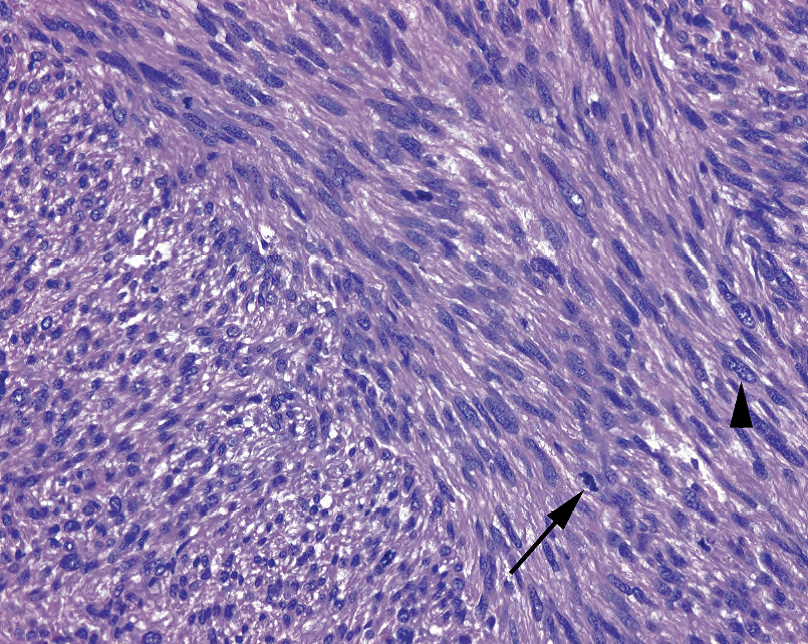
Figure 28.14. Leiomyosarcoma. A malignant version of the leiomyoma, this tumor has the architectural pattern and nuclear morphology of its benign cousin but with much higher cellularity, hyperchromatic nuclei, frequent mitoses (arrow), and large atypical cells (arrowhead).
图28.14 平滑肌肉瘤。与平滑肌瘤对应的恶性肿瘤,具有平滑肌瘤的结构模式和核形态,但细胞密度更高,核深染,核分裂频繁(箭)和非典型大细胞(箭头)。
In a smooth muscle–like lesion arising anywhere near the gastrointestinal tract, you should consider the gastrointestinal stromal tumor (GIST) in the differential diagnosis. Many of what were once called gastric leiomyomas are now identified as GISTS. The cells differentiate along the line of the interstitial cell of Cajal, the pacemaker cell of the stomach, and like this cell, the GIST stains for c-kit and CD34. The GIST may take a spindle cell morphology, overlapping with leiomyoma or schwannoma, or may be epithelioid with a wide range of morphology. Clinical behaviors range from benign to malignant, depending on site and histologic factors.
在胃肠道附近的平滑肌样病变中,应考虑胃肠道间质瘤(GIST)的鉴别诊断。许多曾经被称为胃平滑肌瘤的肿瘤现在被确认为GIST。这些细胞沿着Cajal间质细胞(胃的起搏细胞)的细胞线分化,与此细胞一样,GIST免疫染色显示c-kit和CD34。GIST可能呈梭形细胞形态,与平滑肌瘤或神经鞘瘤重叠,也可能为上皮样细胞,形态多样。根据部位和组织学因素,临床表现从良性到恶性不等。
来源:
The Practice of Surgical Pathology:A Beginner’s Guide to the Diagnostic Process
外科病理学实践:诊断过程的初学者指南
Diana Weedman Molavi, MD, PhD
Sinai Hospital, Baltimore, Maryland
ISBN: 978-0-387-74485-8 e-ISBN: 978-0-387-74486-5
Library of Congress Control Number: 2007932936
© 2008 Springer Science+Business Media, LLC
仅供学习交流,不得用于其他任何途径。如有侵权,请联系删除
本站欢迎原创文章投稿,来稿一经采用稿酬从优,投稿邮箱tougao@ipathology.com.cn
相关阅读
 数据加载中
数据加载中
我要评论

热点导读
-

淋巴瘤诊断中CD30检测那些事(五)
强子 华夏病理2022-06-02 -

【以例学病】肺结节状淋巴组织增生
华夏病理 华夏病理2022-05-31 -

这不是演习-一例穿刺活检的艰难诊断路
强子 华夏病理2022-05-26 -

黏液性血性胸水一例技术处理及诊断经验分享
华夏病理 华夏病理2022-05-25 -

中老年女性,怎么突发喘气困难?低度恶性纤维/肌纤维母细胞性肉瘤一例
华夏病理 华夏病理2022-05-07


共0条评论