外科病理学实践:诊断过程的初学者指南 | 第27章 皮肤
第27章 皮肤(Skin)
The discussion of the skin will be divided into three subsections: melanocytic lesions, nonmelanocytic lesions, and inflammatory (systemic) disorders. Skin biopsies are usually performed because the clinician sees a lesion, such as a mass, a rash, or a macule. However, skin biopsies are also sometimes used to diagnose systemic conditions. Usually the history is enough to direct you to one of the major three categories. Inflammatory skin conditions are not usually diagnosed by the general surgical pathologist, but a working knowledge of their classification can be very helpful. Melanocytic lesions are also more and more the exclusive domain of dermatopathologists, but any surgical pathologist should at least be able to tackle the most benign and most malignant ends of the spectrum.
皮肤的讨论分三节:黑色素细胞病变、非黑色素细胞病变和炎症性(全身性)疾病。通常是因为临床医生看到病变(如肿块、皮疹或黄斑)而进行皮肤活检。然而,皮肤活检有时也用于诊断系统性疾病。病史通常足以引导你进入三大类别之一。炎症性皮肤病通常不是由大外科病理医生来诊断的,但对其分类的工作知识可能非常有用。黑色素细胞病变也越来越成为皮肤病理医生的专属领域,但任何外科病理医生至少应该能够处理这个谱系中最良性和最恶性的两端。
(译注:病理习惯称为黑色素、黑色素细胞、黑色素瘤(MM),皮肤科习惯称为黑素和黑素细胞、黑素瘤)
The grossing of skin biopsy specimens varies a bit by the shape, size, and purpose of the excision, but for diagnostic specimens of tumors, the margins must be entirely examined in perpendicular cuts. See your grossing manual, and consult with your attending, for the best way to cut in a specimen.
皮肤活检标本的大体检查和取材因切除的形状、大小和目的略有不同,但对于肿瘤的诊断标本,必须垂直切开,对切缘进行全面检查。请参阅你的取材手册,并请教你的主治医师,以了解切开样本和取材的最佳方法。
黑色素细胞病变(Melanocytic Lesions)
Melanocytes are specialized cells in the epidermis and elsewhere that are derived from neural crest cells. They have a neuralish, dendritic morphology and stain with S100, like peripheral nerve cells. They also produce melanin pigment, which is exported from the cell and taken up by surrounding epidermal cells. Normal melanocytes do not have much visible pigment; in fact, the cytoplasm is clear, as the pigment leaves the cell (Figure 27.1). Densely pigmented cells along the basal layer of the epidermis are usually basal keratinocytes, not melanocytes. It is this pigment distribution that creates shades of skin color.
黑色素细胞是表皮和其他部位的特殊细胞,起源于神经嵴细胞。它们具有神经化、树突形态和S100染色,就像周围神经细胞。它们还产生黑色素,从黑色素细胞中输出并被周围的表皮细胞摄取。正常黑色素细胞没有太多可见的色素;事实上,细胞质是透明的,因为当色素离开了细胞(图27.1)。沿表皮基底层排列的含有致密色素的细胞通常是基底层角质细胞,而不是黑色素细胞。正是这种色素分布形成了肤色。
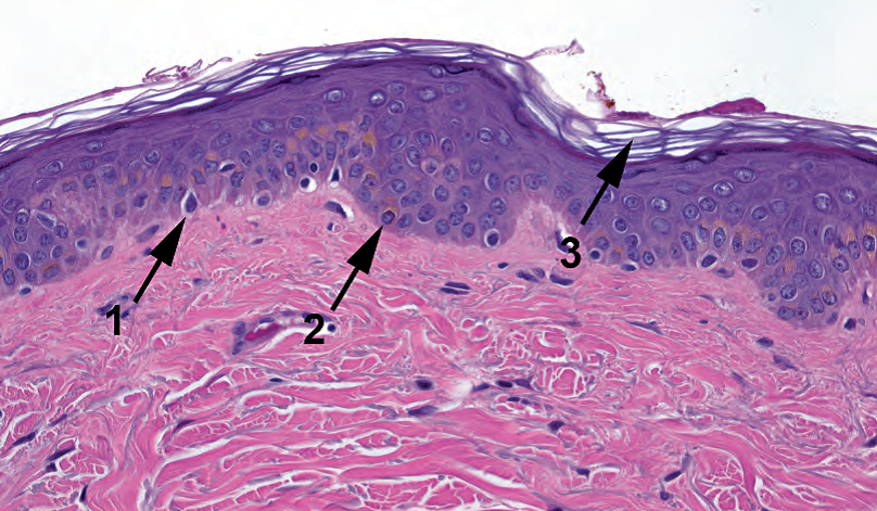
Figure 27.1. Normal melanocyte and skin. A normal melanocyte (1) stands out within a clear halo of cytoplasm. The pigmented component of the skin is actually the basal keratinocytes (2), which absorb the melanin. Typical basket weave or orthokeratin is present (3).
图27.1 正常黑色素细胞和皮肤。一个正常的黑色素细胞(1),细胞质具有透明空晕,显得与众不同。皮肤的色素成分实际上是基底层角质细胞(2),它吸收黑色素。存在典型的角质层(篮网或正角蛋白)(3)。
Abnormal melanocytes can accumulate pigment, and this can be a useful clue in identifying dysplastic melanocytes (discussed below) or identifying an unknown metastasis as melanoma. However, there are plenty of melanomas with no melanin to be found, so do not rely on that. Also beware the melanophage, spindly macrophages in the dermis full of chunky globs of melanin—they are eating it, not making it (Figure 27.2).
异常黑色素细胞可积聚色素,这可能是鉴别异型增生黑色素细胞(下文讨论)或识别未知转移性肿瘤为黑色素瘤的有用线索。然而,很多黑色素瘤没有发现黑色素,所以不要太依赖它。还要注意噬黑色素细胞,即真皮中充满粗大球形黑色素的梭形巨噬细胞,它们吞噬黑色素,而不是制造黑色素(图27.2)。
(译注:dysplastic按病理习惯译为异型增生,皮肤科习惯译为发育不良)
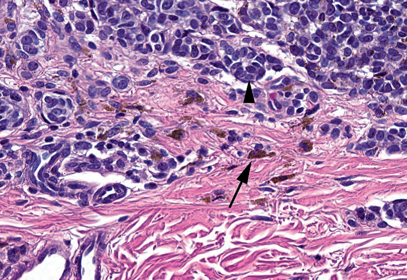
Figure 27.2. Melanophages in an intradermal nevus. The nevus cells (arrowhead) have focal small specks of pigment, but the macrophages digesting the excess melanin (arrow) stand out.
图27.2 皮内痣中的噬黑色素细胞。痣细胞(箭头)有局部小斑点状黑色素,但消化黑色素的巨噬细胞(箭头)显得与众不同。
Become familiar with the melanocyte; spend a few seconds looking for them when you encounter normal skin. Melanoma is a treacherous area precisely because there are no strict diagnostic rules about when something is malignant and when it is not, and much of the diagnosis (in subtle cases) relies on recognizing atypical melanocytes that are up to no good. The only way to learn this skill is to see lots of normal melanocytes.
熟悉黑色素细胞;当你遇到正常皮肤时,花几秒钟寻找它们。黑色素瘤是一个危险的领域,正是因为没有严格的诊断规则来判断某些东西什么时候是恶性的,什么时候又不是恶性的,而且很多诊断(在微妙的情况下)依赖于识别达到不善良程度的非典型黑色素细胞。学习这项技能的唯一方法是多看大量的正常黑色素细胞。
术语(Terminology)
As a pathologist, you now know that a “mole” is a type of gestational trophoblastic disease, and you are sophisticated enough to refer to a bump on the skin as a nevus. However, the word nevus really does mean just a bump on the skin, and there are things called nevus that have nothing to do with melanocytes. In this chapter, we will just be discussing the melanocytic nevi.
作为一名病理医生,你现在知道“mole”表示葡萄胎的时候是一种妊娠滋养层疾病,当你有了一定经验,又会知道“mole”表示皮肤上的一种肿块或隆起的时候,称为痣。然而,“痣”这个词实际上只是指皮肤上的一个肿块,有些叫做痣的东西与黑色素细胞无关。我们在本章只讨论黑色素细胞痣。
A melanocytic nevus is a proliferation of benign melanocytes. It begins along the basal layer of the epidermis, where melanocytes live, and the very earliest manifestation of this is an increased number of melanocytes along the dermoepidermal junction (DEJ) in a single layer. This produces a dark patch on the skin, and the lesion is called a lentigo simplex (Figure 27.3). The word lentigo or lentiginous refers to “along the DEJ” and is used in several different contexts.
黑色素细胞痣是良性黑色素细胞的增殖。正常情况下,黑色素细胞位于表皮的基底层,痣的最早表现是沿着单层的真皮表皮交界处(DEJ)的黑色素细胞数量增多。这会在皮肤上产生一个暗斑,这种病变称为单纯性雀斑(lentigo simplex,图27.3)。
(译注:lentigo原意是“沿着DEJ”,simplex原意是“简单”,合在一起意思是“黑色素细胞简单地沿着DEJ增殖”)

Figure 27.3. Lentigo simplex in acral skin. The dense keratin seen here is typical of acral skin (hands and feet). There is a linear proliferation of single benign melanocytes along the dermoepidermal junction (arrows).
图27.3 肢端皮肤单纯性雀斑。这里所见的致密角蛋白是典型的肢端皮肤(手和脚)。真皮表皮交界处有单个良性黑色素细胞呈线性增殖(箭)。
The next step in the life cycle of a nevus is the proliferation of melanocytes into little nests, or theques, along the DEJ. These are technically intraepidermal, although it is sometimes hard to appreciate that. This lesion is called a junctional nevus, and it appears as little clusters of bland melanocytes hanging from the DEJ (Figure 27.4).
痣生命周期的下一步是黑色素细胞沿着DEJ增殖形成小巢或痣细胞团。从技术上讲,这些是位于表皮内的,但有时很难辨认。这种病变称为交界痣,看似挂在DEJ上的一小簇形态学温和的黑色素细胞(图27.4)。
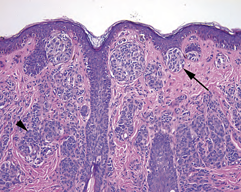
Figure 27.4. Compound nevus. This nevus shows nests of nevocellular (melanocytic) cells attached to the dermoepidermal junction (arrow). A nevus with only dermoepidermal junction nests would be a junctional nevus. In this example, as there are also nevus cells dropping down into the dermis (arrowhead); this is a compound nevus. In a compound nevus, the cells at the deepest point should appear slightly smaller and more bland than those at the dermoepidermal junction (“maturation”).
图27.4 复合痣。此痣显示附在DEJ上的痣细胞(黑色素细胞)巢(箭)。只有DEJ巢的痣是交界痣。本例也有痣细胞像泪滴样向下进入真皮(箭头);这就成为复合性痣。在复合性痣中,最深处的细胞应该比DEJ处的细胞显得更小、形态学更温和(“成熟”)。
From there, the melanocytes may begin to proliferate down into the dermis. They do so as small nests, sheets, or single cells, and they grow in a lobular pattern. Cytologically they are bland, round, clear cells, and they tend to “mature” (become smaller and more bland) the deeper into the dermis they progress. They become so numerous that they make a little nodule in the skin, forming the classic “mole” a la Cindy Crawford. Most adults have 10–20 of them. A nevus with a dermal component plus a junctional component is called a compound nevus. Eventually, with age, the junctional component regresses and you are left with just an intradermal nevus (Figure 27.5). These can be pedunculated, hyperkeratotic, hair bearing, and so forth. Note that melanoma arising in a benign intradermal nevus is vanishingly rare.
从那里,黑色素细胞可能开始增殖并向下进入真皮。它们呈现小巢、成片或单个细胞的形式,并以小叶状模式生长。细胞学温和,呈圆形的透明细胞,进入真皮越深就越“成熟”(变小、形态学更温和)。它们数量如此之多,以至于在皮肤上形成了一个小结节,就像Cindy Crawford那样经典的痣。大多数成人有10-20个痣。兼有真皮成分和DEJ成分的痣称为复合痣。最终,随着年龄增长,DEJ成分退化,只剩下皮内痣(图27.5)。皮内痣可以有蒂、角化过度、有毛发……等等。注意良性皮内痣发生的黑色素瘤是少之又少的。
(译注:Cindy Crawford,美国超级名模,演员,她有标志性美人痣)

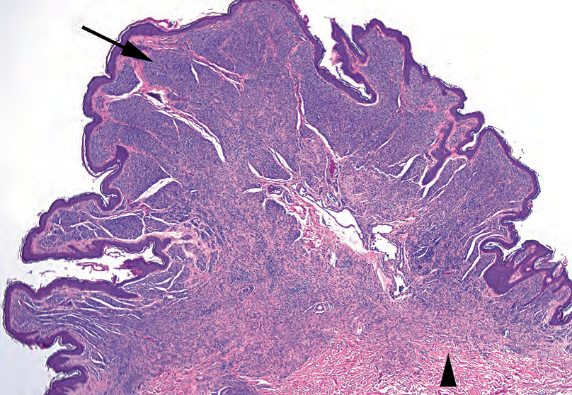
Figure 27.5. Intradermal nevus. This exophytic nevus has only dermal nests of nevus cells (arrow). The lesion is roughly symmetric, and the cells are smaller and more mature at the base (arrowhead).
图27.5 皮内痣。这种外生性痣只有真皮内痣细胞巢(箭)。病变大致对称,底部的细胞更小、更成熟(箭头)。
All of these phases of the nevus share some histologic features of benignity:
痣的所有这些阶段都有一些共同的、良性的组织学特征:
Symmetry
对称性
Size < 3 mm in diameter
直径<3毫米
Lateral borders consisting of nests, not individual trailing melanocytes
由痣细胞巢组成的侧边,而不是单个尾随的黑色素细胞
Lack of atypia in the melanocytes (nuclei are no larger than a keratinocyte nucleus and have small dense nucleoli, if any; multiple nuclei are okay)
黑色素细胞缺乏异型性(核不大于角质细胞核,有小而深染的核仁,如有;多核也可以)
Maturation into the dermis
进入真皮后变得更成熟
Chunky brown-black pigment
色素呈粗大的棕黑色
(译注:皮内痣是“位于真皮内的痣”。交界痣是“位于真皮表皮交界处的痣”,并非病变性质是交界性。兼有两种痣,则为复合痣)
其他良性痣(Other Benign Nevi)
The common blue nevus consists of a diffuse scattering of pigmented, dendritic (stellate), single melanocytes in the dermis (Figure 27.6). They are mixed in with melanophages.
普通蓝痣由真皮中弥漫分布的散在的含有色素的树突状(星芒状)单黑个色素细胞组成(图27.6),夹杂着噬黑色素细胞。
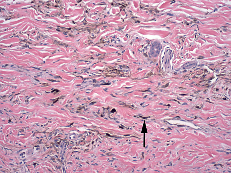
Figure 27.6. Blue nevus. Small, indistinct, pigmented cells are scattered throughout the dermal collagen (arrow). The cells are elongated and fusiform or stellate and do not make rounded nests like typical nevus cells. Some of the larger cells with chunky pigment are likely melanophages.
图27.6 蓝色痣。小的、不清楚的、含色素的细胞,散布在整个真皮胶原中(箭)。细胞被拉长,呈梭形或星芒状,不像典型的痣细胞那样形成圆形细胞巢。一些较大的细胞含有粗糙色素,可能是噬黑色素细胞。
The Spitz nevus is usually found on the head and neck of children and adolescents. At low power it is circumscribed and symmetric, and large nests of melanocytes are found between skinny elongated rete (Figure 27.7). Eosinophilic Kamino bodies may be seen at the DEJ. The reason this lesion is so troublesome is that the melanocytes may be large, spindled, pleomorphic, or atypical, and they may even show rare mitoses, which suggests melanoma.
Spitz痣通常见于儿童和青少年的头颈部。低倍镜下有边界,对称,在拉长的上皮脚之间发现黑色素细胞形成的大巢(图27.7)。DEJ处可见嗜酸性Kamino小体。这种病变之所以如此麻烦,是因为黑色素细胞可能增大、梭状、多形性或非典型,甚至可能有少量核分裂,这提示黑色素瘤。
(译注:Kamino小体(嗜酸性小体,KB)是表皮内粉红色圆形小体,边缘不规则。KB最初是在Spitz痣中描述的;后来也有报道称其存在于其他病变中,如Reed痣、Spitz样肿瘤和恶性黑色素瘤。Kamino等人研究表明,表皮中PAS和三色染色阳性的KB有助于区分Spitz痣与恶性黑色素瘤。详见https://www.ncbi.nlm.nih.gov/pmc/articles/PMC6503810/)
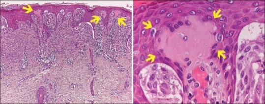
HE染色示Spitz痣中的Kamino小体(嗜酸性小体,KB)
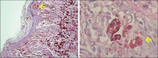
PAS染色示Spitz痣中的Kamino小体(嗜酸性小体,KB)
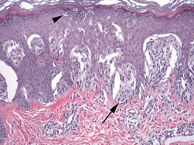
Figure 27.7. Spitz nevus. This nevus in a child shows nests of large, spindly melanocytes at the dermoepidermal junction (arrow) and rare melanocytes spreading up through the epidermis (arrowhead). In an adult, this pattern would be very worrisome.
图27.7 Spitz痣。儿童的痣,显示DEJ处有大的梭形黑色素细胞形成的细胞巢(箭),少数黑色素细胞通过表皮向上扩散(箭头)。如果是成年人的痣,这种模式非常令人担忧。
Acral and genital nevi—nevi of the hands and feet, genital regions, and breasts—are allowed some atypical features. These nevi have prominent lentiginous growth, with occasional ascending cells mimicking pagetoid spread. However, they should not have cytologic atypia.
肢端和生殖器痣—手足、生殖器区域和乳房的痣,可以有一些非典型特征。这些痣有明显的雀斑样生长,偶尔有向上的细胞貌似Paget样扩散。然而,他们不应该有细胞学非典型。
Most nevi are acquired during childhood to early adulthood, but some are congenital. To have congenital features means that the dermal melanocytes tend to track down the adnexal structures.
大多数痣是获得性,在儿童期至成年早期发生;但有些是先天性。具有先天特征意味着真皮黑色素细胞倾向于沿着皮肤附属器结构向下生长。
异型增生痣(Dysplastic nevi)
There are some nevi that begin to show some features more commonly associated with melanoma. These nevi are clinically distinct looking, and although they are not considered actual precursors to melanoma, patients with multiple dysplastic nevi are at significantly higher risk of developing melanoma. However, “dysplastic nevus” is a clinical diagnosis, and as pathologists we merely describe the features we see. There are two components to dysplasia in this context: architectural disorder and atypia. These lesions are signed out as, for example, Compound nevus with architectural disorder and severe cytologic atypia. (However, in some texts you will find this entity listed as “lentiginous melanocytic nevus.”)
有些痣开始表现出一些与黑色素瘤相关的特征。这些痣在临床上看起来很独特,虽然它们不是黑色素瘤的实际前驱病变,但患有多发性异型增生痣的患者,黑色素瘤的风险明显较高。然而,“异型增生痣”是一种临床诊断,作为病理医生,我们只描述我们所看到的特征。在这种情况下,异型增生有两个组成部分:结构紊乱和细胞非典型。例如可以这样签发报告:复合痣伴结构紊乱和重度细胞非典型。(然而,在一些文章中,你会发现这个实体被列为“雀斑样黑色素细胞痣”。)
There are four features of architectural disorder: Architectural disorder is not graded but simply present or absent.
结构紊乱不用分级,只要注明有或无。结构紊乱有四个特征:
Lentiginous spread of atypical melanocytes (along the DEJ in a creeping line)
非典型黑色素细胞的雀斑样扩散(沿着DEJ爬行,线性排列)
Shouldering (the lentiginous component is wider than the dermal component)
肩带现象(雀斑样成分比真皮成分宽)
Bridging of rete (nests attached to adjacent rete ridges fuse; Figure 27.8)
上皮脚相连(附在相邻上皮脚上的痣细胞巢相互融合;图27.8)
Fibroplasia (a feathering of the dermal collagen that looks like pink cotton candy)
纤维化(真皮胶原的羽毛状物,看起来像粉红色的棉花糖)
The features of cytologic atypia include the following:
细胞学非典型的特征包括:
Hyperchromatic nuclei, increased nuclear to cytoplasmic ratio
核深染,核质比增加
Large red nucleoli
大红核仁
Accumulation of dusty grey-brown melanin (see Figure 27.8)
粉尘状灰棕色黑色素的积聚(见图27.8)
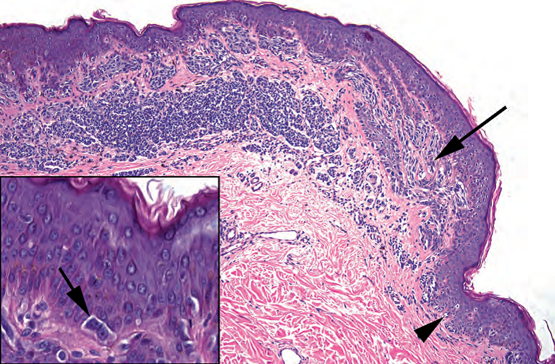
Figure 27.8. Dysplastic nevus. At low power, elongated nests of spindly melanocytes are seen bridging across adjacent rete (arrow), and single melanocytes trail off to the lateral edge of the lesion (arrowhead). These are features of architectural disorder. Inset: Atypical melanocytes with large nuclei and nucleoli are seen at the dermoepidermal junction.
图27.8 异型增生痣。在低倍镜下,拉长的黑色素细胞巢桥接相邻的上皮脚(箭),单个黑色素细胞沿着病变的外侧边缘延伸(箭头)。这些都是结构紊乱的特征。插图:DEJ处可见非典型黑色素细胞,核增大,可见核仁。
Atypical mitoses
非典型核分裂
Atypia is graded as mild, focally severe, or severe.
细胞非典型分为轻度、局灶性重度或重度。
In general, these nevi tend to be suspicious enough that you must take a few moments to prove to yourself that they are not melanoma (see next section).
总的来说,这些痣往往很可疑,你必须花一些时间来说服自己,它们不是黑色素瘤(见下一节)。
黑色素瘤(Melanoma)
The best way to think about melanoma is as the presence of malignant melanocytes. Because melanocytes can proliferate in many ways and still be benign, it takes considerable experience to decide if a melanocyte is malignant or not. However, setting that aside for a moment, the types of melanoma include the following:
考虑黑色素瘤的最佳方式是存在恶性黑色素细胞。因为黑色素细胞可以以多种方式增殖,并且仍然是良性,所以判断黑色素细胞是否恶性,需要相当多的经验。然而,暂且不提这一点,黑色素瘤的类型包括:
Lentigo maligna: malignant melanocytes proliferating only along the DEJ
恶性雀斑:只有沿着DEJ增殖的恶性黑色素细胞
Melanoma in situ: malignant melanocytes along the DEJ, and percolating up through the epidermis in a pagetoid fashion (something benign melanocytes just do not do)
原位黑色素瘤:沿着DEJ的恶性黑色素细胞,以Paget样方式向上渗入表皮(良性黑色素细胞不会这样)
Malignant melanoma: malignant melanocytes along the DEJ, pageting through the epidermis and invading the dermis
恶性黑色素瘤:沿着DEJ的恶性黑色素细胞,以Paget样方式向上浸润表皮,向下浸润真皮
Superficial spreading melanoma: melanoma in a “horizontal growth phase,” meaning it is spreading laterally along the DEJ but also involves the dermis (clinically, this is a macular lesion [flat])
浅表扩散性黑色素瘤:处于“水平生长期”的黑色素瘤,这意味着它沿着DEJ横向扩散,但也累及真皮(临床上,这是一种黄斑病变[平坦])
Nodular melanoma: melanoma with a “vertical growth phase,” meaning that it is primarily growing down into the dermis (almost like an intradermal nevus, but with malignant cells) and that it produces a raised lesion
结节性黑色素瘤:具有“垂直生长期”的黑色素瘤,这意味着它主要向下生长进入真皮(几乎像皮内痣,但有恶性细胞),并产生隆起的病变
Most melanomas have both a horizontal and a vertical component, which is the classic irregularly shaped dark macule with a central raised or ulcerated papule.
大多数黑色素瘤兼有水平成分和垂直成分,呈典型的不规则形状的暗斑,中央隆起或溃疡性丘疹。
恶性特征(Features of Malignancy)
Unfortunately there is no single feature than can rule melanoma in or out. As with many types of neoplasia, there are certain features that suggest malignancy, and the presence of enough of them can convince you of the diagnosis. Many of these criteria are subjective and require experience, which is why dermatopathology is such a booming subspecialty these days. On low power, look for the following:
不幸的是,没有单个特征可以诊断或排除黑色素瘤。正如其他多种肿瘤,有些特征提示恶性,足够多的特征可能使你确定诊断。这些标准中有许多是主观的,需要经验,这就是皮肤病理学现在蓬勃发展的原因。在低倍镜下,寻找以下各项:
Asymmetry
不对称
Poorly circumscribed, pleomorphic, discohesive nests of melanocytes
黑色素细胞巢边界不清、多形性、不粘附
Shouldering (lateral spread) of atypical melanocytes
非典型黑色素细胞的肩带现象(侧向扩散)
Pagetoid spread through the epidermis (Figure 27.9)
Paget样扩散进入表皮(图27.9)
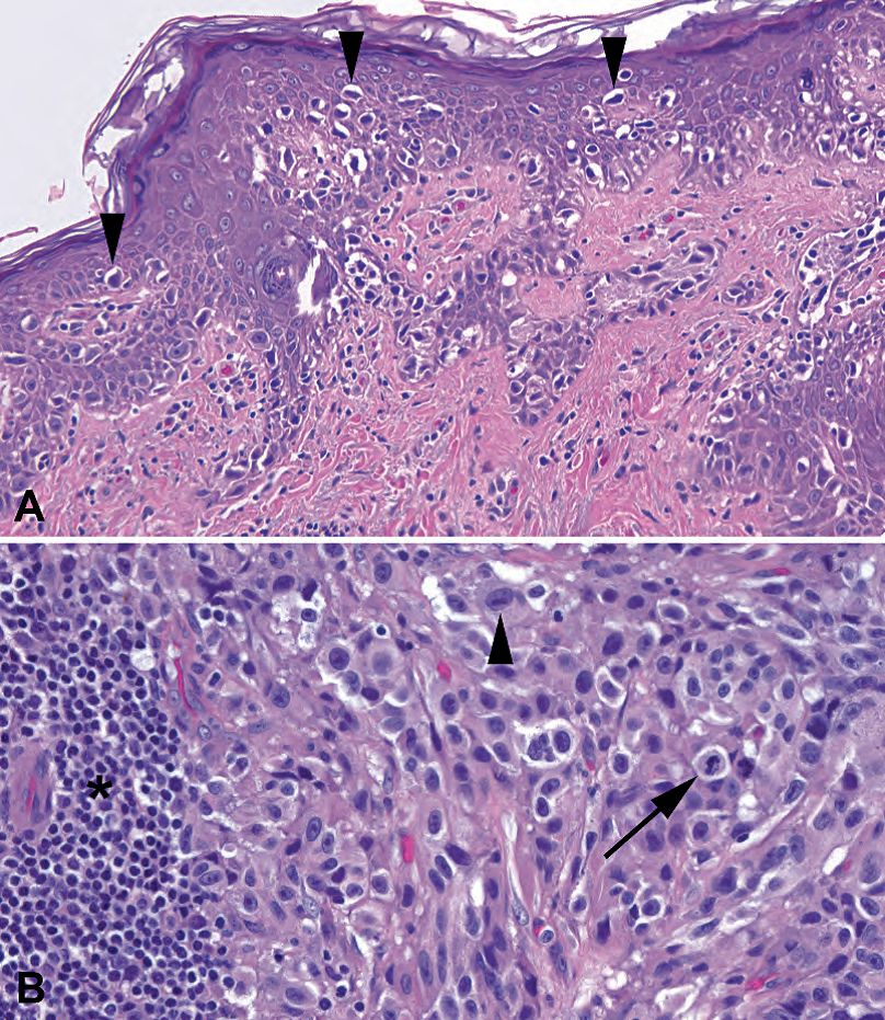
Figure 27.9. Melanoma. (A) Large, dark, irregular melanocytes can be seen infiltrating upward through the epidermis (arrowheads) in pagetoid fashion. If there were no dermal component, this would qualify as melanoma in situ. (B) Malignant melanocytes deep in the dermis. Large atypical nuclei with large nucleoli (arrowhead) plus the presence of mitoses (arrow) is diagnostic of invasive melanoma. Adjacent lymphocytes (asterisk left side of the field) are common.
图27.9 黑色素瘤。(A)大的、深染的、不规则的黑色素细胞,以Paget样方式向上浸润表皮(箭头)。如果没有真皮成分,这定性为原位黑色素瘤。(B)深入真皮的恶性黑色素细胞。大的非典型核和大核仁(箭头)加上核分裂(箭)是浸润性黑色素瘤的诊断特征。常见相邻的淋巴细胞(视野左侧的星号)。
Associated lymphocytes, especially band-like
相关淋巴细胞,尤其是带状淋巴细胞
On high power, look for the following:
在高倍镜下,寻找以下各项:
Atypia, as described earlier
非典型,如前所述
Lack of deep maturation (for dermal component)
缺乏深部成熟(真皮成分)
Mitoses or atypia in the dermis (see Figure 27.9)
真皮成分的核分裂或异型性(见图27.9)
Melanocytic necrosis
黑色素细胞坏死
签发报告的标准(Sign Out Criteria)
All diagnoses of invasive (into the dermis) melanoma must include certain prognostic features. The first is the depth of invasion. This is called Breslow’s thickness and is the depth (to the hundredth of a millimeter) of invasion from the top of the epidermal granular layer to the deepest malignant cell. It is functionally equivalent to stage; the deeper the invasion, the poorer the prognosis. Clark’s level is a related concept but is based on the histologic layers or levels of the dermis, not the absolute depth.
所有浸润性(进入真皮)黑色素瘤的诊断必须包括某些预后特征。首先是浸润深度。这称为Breslow厚度,是从表皮颗粒层的顶部到最深部恶性细胞的浸润深度(精确到百分之一毫米)。它在功能上等同于分期;浸润越深,预后越差。Clark水平是一个相关的概念,但基于真皮的组织学层次或水平,而不是绝对深度。
The second important prognostic feature is the presence or absence of ulceration. The third is the margin status, both deep and lateral.
第二个重要的预后特征是有无溃疡。第三是切缘状态,包括深切缘和侧切缘。
黑色素瘤的特殊类型(Special Types of Melanoma)
In desmoplastic and spindle cell melanoma, melanocytes can become very spindly and sarcomatoid. With an unidentified spindle cell lesion in the dermis, you must always rule out melanoma. The scariest and most subtle form is the desmoplastic melanoma, which is not only spindly but often sparsely cellular in a background of dense collagen; in other words, it looks just like a scar. A useful tip-off to a lurking desmoplastic melanoma is, aside from a slightly “busy” dermis, the presence of bands or clumps of lymphocytes (Figure 27.10).
促结缔组织增生性黑色素瘤和梭形细胞黑色素瘤,黑色素细胞可以变成非常明显的梭形和肉瘤样。真皮中的不明梭形细胞病变,必须排除黑色素瘤。最恐怖和最微妙的形式是促结缔组织增生性黑色素瘤,它不仅梭形,而且通常是细胞稀疏,背景是密集的胶原蛋白;换句话说,它看起来就像瘢痕。隐匿性促结缔组织增生性黑色素瘤的一个有用提示是,除了有点紊乱的真皮外,还存在淋巴细胞带或淋巴细胞团(图27.10)。
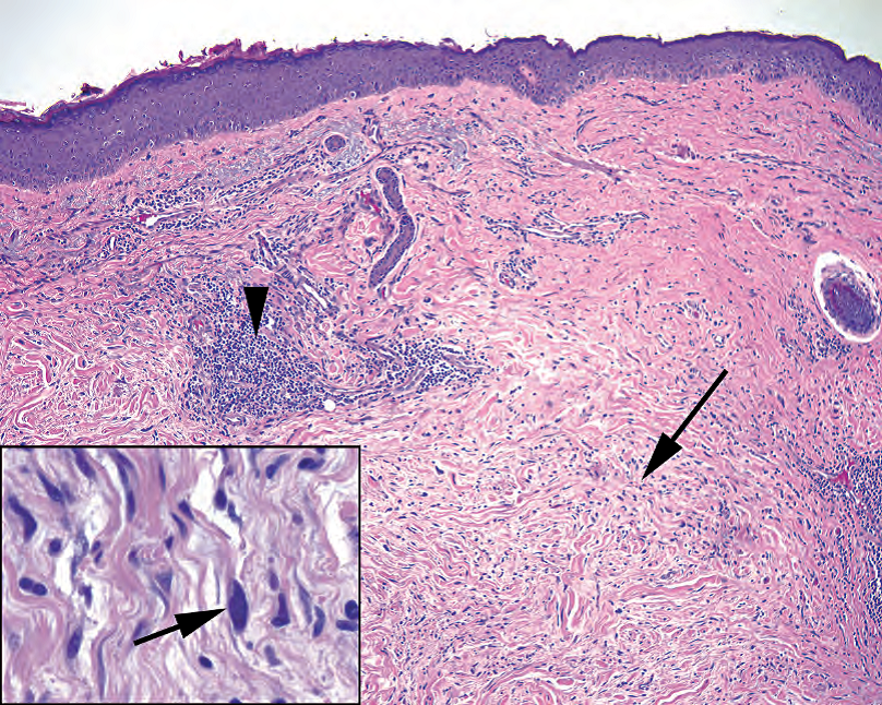
Figure 27.10. Desmoplastic melanoma. At low power, there appears to be a hypocellular scar in the dermis (arrow). The clue to melanoma lies in the collection of lymphocytes (arrowhead). Inset: On higher power, there are enlarged and hyperchromatic cells (arrow) in the “scar.” These would be positive for S100, unlike fibroblasts.
图27.10 促结缔组织增生性黑色素瘤。低倍镜下,真皮中似乎有一个细胞稀少的瘢痕(箭)。黑色素瘤的线索在于淋巴细胞的聚集(箭头)。插图:在高倍镜下,“瘢痕”中有增大的深染的细胞(箭)。这些细胞呈S100阳性,与成纤维细胞不同。
Another type of melanoma is acral lentiginous. Acral refers to the distal extremities. Like the benign acral nevus, this lesion is characterized by prominent lentiginous growth. It can be very difficult to distinguish from an acral nevus.
另一种黑色素瘤是肢端雀斑样黑色素瘤。肢端是指远端肢体。与良性肢端痣一样,该病变的特征是显著的雀斑样生长。它很难区分肢端痣。
Metastases can look like anything at all; they can be spindly, epithelioid, rhabdoid, small cell, and so forth. However, common features of metastases include alveolar (nested) architecture, large pink-to-violet cells with big nuclei and red nucleoli, and occasional melanin pigment. As a rule of thumb, if you don’t know what it is, consider melanoma.
转移性黑色素瘤可能看似任何东西;它们可以梭形、上皮样、横纹肌样、小细胞……等等。然而,转移的共同特征包括腺泡状(巢状)结构,大的粉红色到紫色细胞,有大核和红核仁,偶尔还有黑色素。作为一个经验法则,如果你不知道它是什么,考虑黑色素瘤。(译注:看到四不象,考虑MM;鉴别诊断永远记得MM)
黑色素瘤的再切除术(Re-excision of Melanomas)
When a melanoma is diagnosed on excisional biopsy, it is nearly always given a wide re-excision. You will see these huge ellipses on surgical pathology. Pathologists differ in how much of the re-excision to submit, but at the least the entire biopsy scar, to the lateral margins, should be submitted. Carefully scan not just the epidermis but also the dermis deep to the biopsy site.
切除活检诊断黑色素瘤后,几乎总是扩大切除(广泛的再次切除)。你会在外科病理取材室看到这些巨大的椭圆形皮肤标本。取材数量存在分歧,但至少应切取整个活检瘢痕至现两侧切缘。镜下仔细扫描,不仅观察表皮,还有活检部位深部的真皮。
免疫组化染色(Special Stains)
S100 is the workhorse stain for melanoma, as it stains all types. HMB-45 and Melan-A are also melanoma markers, but notably do not stain spindled or desmoplastic melanomas. Also remember that there is a whole family of tumors that are HMB-45 positive but not melanoma (angiomyolipoma and the perivascular epithelioid cell tumors).
S100是黑色素瘤的主要抗体,因为它能染所有类型的黑色素瘤。HMB-45和Melan-A也是黑色素瘤标志物,但不染色梭形细胞或促结缔组织增生性黑色素瘤。还要记住,HMB-45阳性肿瘤是一个完整的家族,不止是黑色素瘤,如:血管平滑肌脂肪瘤、和血管周围上皮样细胞瘤(PEComa)。
(译注:HMB-45阳性肿瘤还有淋巴管平滑肌瘤病和肺的“糖瘤”。Melan-A也能染肾上腺的皮质癌和性腺的性索肿瘤)
Sentinel nodes for melanoma, if negative by H&E, are stained using HMB-45 and MelanA. There are inherent S100-positive cells in lymph nodes, so it is not a good screening stain for this purpose.
黑色素瘤前哨淋巴结,如果HE染色阴性,则使用HMB-45和MelanA染色。淋巴结中存在固有的S100阳性细胞,因此不太好用。
There are, unfortunately, no stains that can differentiate a benign nevus from a malignant melanoma. However, in general, HMB-45 should only stain the most superficial cells in a nevus, as the deeper maturing component loses the antigen. Similarly, Ki-67, a proliferation marker, should only be positive at the surface of a nevus, not deep.
不幸的是,没有哪种染色能区分良性痣和恶性黑色素瘤。然而,总的来说,HMB-45应该只是标记痣中最表浅的细胞,因为更深的成熟成分丢失HMB-45抗原。类似地,Ki-67作为一种增殖标记物,应该只在痣表面呈阳性,而不是深层。
非黑色素细胞病变(Nonmelanocytic Lesions)
This section will address the lesions, both neoplastic and hyperplastic, that are not made of melanocytes. These include squamous lesions, cysts, adnexal tumors, and miscellaneous common soft tissue tumors of the dermis.
本节将讨论不是由黑色素细胞构成的病变,包括肿瘤性病变和增生性病变。这些包括鳞状病变、囊肿、皮肤附属器肿瘤和常见的真皮软组织肿瘤。
日光损伤(Sun Damage)
The first major category of tumors is the spectrum of disease seen in sun-exposed skin, typically the face, neck, and arms of adults. A general marker of sun exposure is solar elastosis, which is an accumulation of grey wispy damaged elastin in the dermis (Figure 27.11). Ironically, it represents a loss of elasticity (wrinkles). One of the benign changes seen in the context of sun exposure is the solar lentigo (lentigo senilis, age spot), which appears as a finger-like proliferation of hyperpigmented rete growing down from the epidermis (Figure 27.12). Keratinocytes, not melanocytes, are the pigmented cells.
第一大类肿瘤是一个疾病谱系,发生在日光暴露的皮肤,通常是成人的面部、颈部和手臂。日光暴露的一个共同标志是日光性弹性纤维变性,它是真皮中累积的灰白色受损的弹性蛋白(图27.11)。心塞的是,它造成皮肤弹性丧失(皱纹)。在日光暴露的情况下,可以看到的良性改变之一是日光性雀斑(老年雀斑),它表现为色素增多的上皮脚,从表皮向下生长呈手指状增殖,(图27.12)。含有色素的细胞是角质细胞,而不是黑色素细胞。
(译注:solar lentigo,又称日光性黑子、日光性小痣、晒斑、太阳痣)
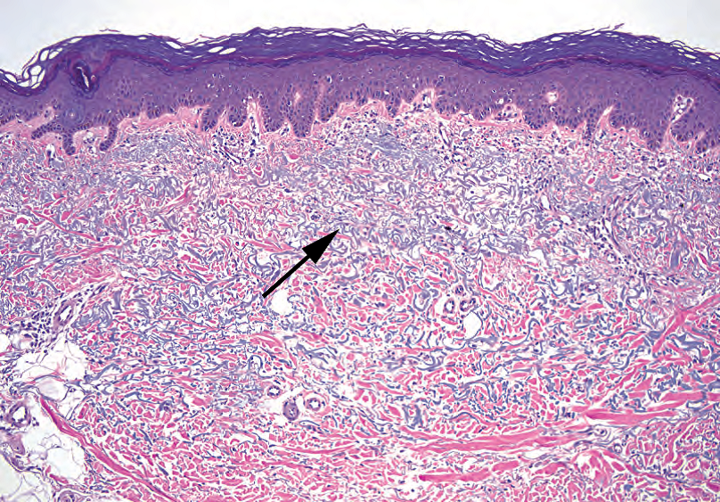
Figure 27.11. Solar elastosis. This is the typical microscopic appearance of sun-damaged skin. The collagen is replaced by wispy gray-blue strands of elastin (arrow).
图27.11 日光性弹性纤维变性。这是日光损伤皮肤的典型镜下表现。胶原被一缕缕纤细的灰蓝色弹性蛋白取代(箭)。
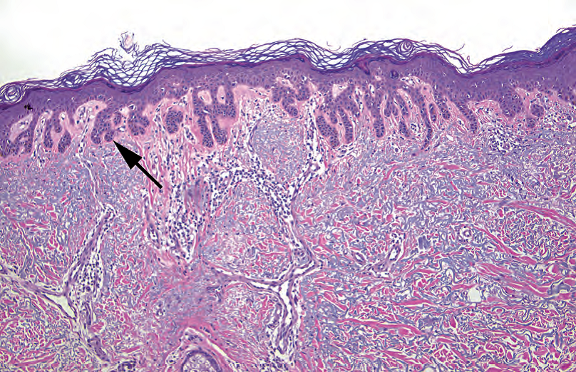
Figure 27.12. Solar lentigo. Prominent rete are growing down from the epidermis (arrow), with increased basal pigmentation (not clearly visible at this power). Notice the underlying solar elastosis. Compare this lesion to the lentigo simplex (see Figure 27.3), which, in contrast, shows a proliferation of melanocytes.
图27.12 日光性雀斑。明显的上皮脚,从表皮向下生长(箭),基底层色素增多(这个放大倍数看不清)。注意下方的日光性弹性纤维变性。与单纯性雀斑相比(见图27.3),后者显示黑色素细胞增殖。
The solar lentigo may then develop into a dysplastic lesion called an actinic keratosis. Actinic keratoses have a wide variety of appearances, from very thin with subtle atypia to very hypertrophic with full-thickness atypia. However, the defining features of an actinic keratosis should include the following:
日光性雀斑随后可能发展成异型增生病变,称为日光性角化病(又称光化性角化病)。日光性角化病有多种表现,从非常薄的轻微非典型到非常显著增生的全层非典型。然而,日光性角化病的定义特征应包括以下内容:
Squamous atypia of varying thickness, often noticeable only in comparison to the surrounding uninvolved epidermis (Figure 27.13)
不同厚度的鳞状细胞非典型,通常只是在对比周围未受累的表皮之后才会觉得明显(图27.13)

Figure 27.13. Actinic keratosis. This example shows an area of disorganized and enlarged nuclei (1) with prominent and atypical mitoses (2), consistent with dysplasia. There is overlying hyperkeratosis and parakeratosis (3) and underlying solar elastosis (4). This is a slightly tangential cut through the skin, making the lesion appear very thick.
图27.13 日光性角化病。这例显示一个区域的核变得无序和增大(1),有明显的核分裂和非典型核分裂(2),这些表现符合异型增生。上方角化过度和角化不全(3),下方日光性弹性纤维变性(4)。这例稍微斜切面,使病变看起来很厚。
Alteration of the keratinization to become pink and parakeratotic
角化变为粉红色和角化不全
Sparing of the keratin above the hair follicles, classically resulting in alternating columns of parakeratosis and orthokeratosis
毛囊上方的角蛋白稀疏,导致典型的角化不全和角化正常的交替排列
Underlying solar elastosis
下方日光性弹性纤维变性
(译注:英文术语医学词典https://medical-dictionary.thefreedictionary.com/
orthokeratosis 正角化或角化正常:表皮的角质层形成一层透明的角蛋白层,如正常表皮。[ortho-正,keras-角,-osis状态]
hyperkeratosis 角化过度:表皮或粘膜的角质层增厚。
parakeratosis 角化不全:一种角化过度模式,角质层仍有核,见于多种鳞屑性皮肤病,如银屑病和亚急性或慢性皮炎。)
Actinic keratosis is regarded, conceptually, as a form of carcinoma in situ, but its natural history is unpredictable. Actinic keratoses can (rarely) invade before they reach full-thickness atypia, unlike squamous lesions of the cervix. However, when the atypia does reach full thickness (assuming no invasion), these lesions may be called bowenoid actinic keratosis or just squamous cell carcinoma in situ to emphasize the severity of the lesion.
日光性角化病在概念上视为是原位癌的一种形式,但其自然史是不可预测的。与宫颈鳞状病变不同,日光性角化病可能(很少)在达到全层非典型之前就发生浸润。然而,当异型性达到全层时(假设没有浸润),这些病变可以称为鲍温样日光性角化病或直接称为原位鳞状细胞癌,以强调病变的严重性。
Carcinoma in situ is a simple concept in other organs, but in the skin it is a source of great debate. Most dermatopathology chapters are littered with the phrase “we use the term…”— meaning each camp has their own philosophy and style. Part of the problem is that the path from dysplasia to invasive carcinoma looks more like a metro subway map than a straight line. However, the basic idea is that carcinoma in situ has not yet crossed the basement membrane and that there is some degree of epidermal dysplasia. Entities that fall under the category of carcinoma in situ include the following:
原位癌在其他器官是简单的概念,在皮肤却是争论的焦点。大多数皮肤病理学专著都声明“我们使用某某术语”,这意味着每个阵营都有自己的哲学和风格。部分问题在于,从异型增生到浸润性癌的路径看起来更像地铁地图,而不是直线。然而,基本观点是原位癌尚未穿透基底膜,并存在一定程度的表皮异型增生。属于原位癌类别的疾病实体包括:
Actinic keratosis (as discussed earlier) and bowenoid actinic keratosis
日光性角化病(如前所述)和鲍温样日光性角化病
Bowen’s disease—often used as a synonym for carcinoma in situ but actually describes a particular clinical presentation that occurs on non–sun-damaged skin and does not spare the hair follicles, unlike the actinic keratosis family
Bowen病,常用作原位癌的同义词,但实际上描述一种特殊的临床表现,发生在未受阳光照射的皮肤,会累及毛囊,这不同于日光性角化病家族
Bowenoid papulosis—a human papillomavirus–related lesion of genital sites
鲍温样丘疹病,与HPV相关的生殖器部位病变
Invasive squamous cell carcinoma is most likely to arise from the sun-damaged, actinic keratosis–type pathway and hence is usually seen in the background of solar elastosis and actinic keratosis–like changes. Features that suggest invasion include penetration of nests deep into the dermis, accompanied by an aberrant deep keratinization (pinkness). Finding single cells invading the dermis is fairly conclusive (Figure 27.14). The appearance of squamous cell carcinoma is similar to that found in other sites (see Chapters 16 and 22).
浸润性鳞状细胞癌最有可能起源于日光损伤的日光性角化病型途径,因此通常出现在日光性弹性纤维变性和日光性角化病样改变的背景下。提示浸润的特征包括细胞巢向下穿透深部真皮,伴有异常的深部角化(粉红色)。发现单个细胞浸润真皮是相当确凿的证据(图27.14)。鳞状细胞癌的表现类似于其他部位(见第16章和第22章)。
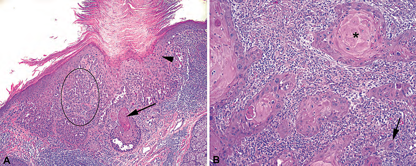
Figure 27.14. Squamous cell carcinoma. (A) Superficially invasive squamous cell carcinoma, showing the paradoxic deep keratinization (arrow), indicating an invasive nest. Actinic keratosis–type changes are seen in the overlying epidermis (arrowhead), including hyperkeratosis. In one area, the pattern of thin cords of cells infiltrating the stroma (oval) is too complex to be explained by a funny plane of section and is another pattern of invasion. (B) Higher power view of invasive squamous cell carcinoma, showing keratin pearls (asterisk) and infiltrating single cells (arrow).
图27.14 鳞状细胞癌。(A)浅表浸润性鳞状细胞癌,表现为反常的深部角化(箭),提示浸润性细胞巢。日光性角化病样改变见于上方表皮(箭头),包括角化过度。在一个区域,浸润间质的模式呈细长条索状(卵圆形)太复杂,无法用有趣的切面原因来解释,这是另一种浸润模式。(B)浸润性鳞状细胞癌的高倍视野,显示角化珠(星号)和单个细胞浸润(箭)。
Basal cell carcinoma is another of the common sun-related tumors. It is the most common cutaneous malignancy and, despite its reputation as a sort of ho-hum and uninteresting tumor, has a very wide range of appearances. There is also some overlap between basal cell carcinoma and benign adnexal tumors; the latter are probably often missed. Features of basal cell carcinoma include the following:
基底细胞癌是另一种常见的日光相关肿瘤。它是(美国)最常见的皮肤恶性肿瘤,尽管认为它是一种令人乏味的、无趣的肿瘤,但它的表现非常广泛。基底细胞癌和良性附属器肿瘤之间也有一些重叠;后者可能经常被忽略。基底细胞癌的特征包括:
Lobules of small, blue, basal-type keratinocytes with peripheral palisading (picket-fence) arrays of oblong nuclei (Figure 27.15)
小蓝色基底型角质细胞形成的小叶结构,周围有栅栏状排列的长圆形细胞核(图27.15)
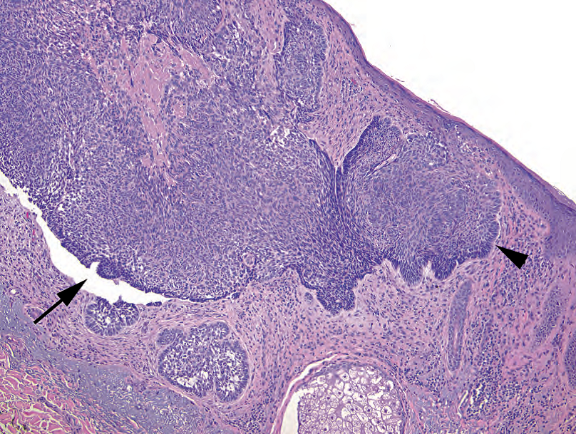
Figure 27.15. Basal cell carcinoma. Blue nests of cells appear to drop down from the epidermis. There is prominent palisading of the basal cells at the periphery of the nests (arrowhead) and clefting of the tumor cells away from the stroma (arrow).
图27.15 基底细胞癌。蓝色的细胞巢似乎从表皮脱落下来。巢周围的基底细胞有明显的栅栏状排列(箭头),肿瘤细胞从间质中撕裂出来(箭头)。
Formation of clefts (cracks) between the tumor nests and the stroma
肿瘤巢和间质之间形成裂隙
Sometimes (not always) desmoplasia, focal keratinization, or mucin production
有时(并非总是)结缔组织增生、局灶性角化或粘液生成
At low power the basal cell carcinoma nests can look similar to adnexal structures, making margins challenging. However, basal cell carcinoma tumor cells should have darker chromatin, more apoptosis and mitoses, and paler cytoplasm than the hair follicles.
在低倍镜下,基底细胞癌巢可能看似附属器结构,难以判断切缘。然而,与毛囊相比,基底细胞癌的染色质较深染,凋亡和核分裂较多,细胞质较淡染。
Special types of basal cell carcinoma include nodular (the usual type), superficial multicentric, and sclerosing. The superficial multicentric form tends to hang off the epidermis like stalactites, without forming a mass, and can have skip areas (harder to excise). The sclerosing form shows a prominent desmoplastic response. There are up to 20 more subtypes; a large dermatopathology atlas will show the many faces of basal cell carcinoma.
特殊类型的基底细胞癌包括结节型(普通型)、浅表多中心型和硬化型。浅表多中心型就像钟乳石一样悬挂在表皮上,不形成肿块,并且可能有跳跃区域(更难切除)。硬化型表现为显著的促结缔组织增生反应。有多达20多个亚型;大本的皮肤病理图谱会显示基底细胞癌的许多面孔。
其他角化过度但非肿瘤性病变(Other Hyperkeratotic but Nonneoplastic Lesions)
Seborrheic keratoses are very common, benign lesions that have many, many forms, but the usual picture is a hyperkeratotic, orthokeratotic lesion with a markedly thickened epidermis. It often forms a raised plaque on the skin; on the slide, the epidermis looks as though it was accidentally cut en face, with convoluted, confluent cords of epidermis. Horn cysts, which are entrapped whorls of orthokeratin, are common (Figure 27.16). (These are quite different from the squamous pearls of carcinoma, which are pink and parakeratotic.) Pigment and inflammation may be seen; atypia is not. These are not necessarily associated with sun damage.
脂溢性角化病是一种很常见的良性病变,有很多种形式,但通常表现为角化过度、正角化的病变,表皮明显增厚。它通常形成皮肤隆起的斑块;镜下,表皮看起来像是意外地在脸上切下的,有卷曲的、融合的表皮条索。常见角囊肿,它是表皮包裹的正角蛋白旋涡(图27.16)。(比较:鳞癌的角化珠呈粉红色,角化不全。)可见色素和炎症;非典型性并不重要。这些不一定与太阳伤害有关。
(译注:脂溢性角化病诊断要点:基底平坦;基底样细胞增生,可有色素;角化过度和角囊肿)
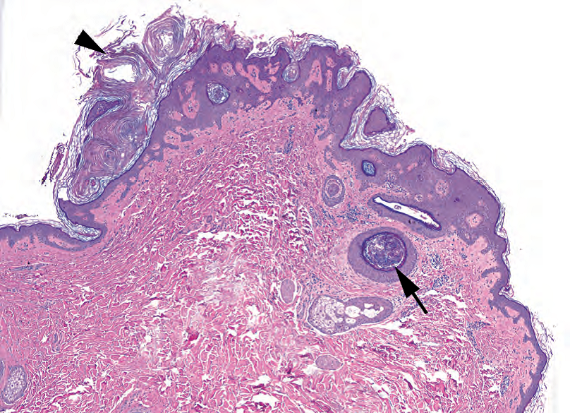
Figure 27.16. Seborrheic keratosis. This exophytic lesion shows hyperkeratosis (arrowhead) but not parakeratosis (no visible nuclei in the keratin). The epidermis takes on a complicated pattern of intertwining rete, and in some areas foci of keratin are trapped within the lesion, forming horn cysts (arrow). Compare these blue, acellular, lamellated balls of keratin with the pink keratin pearls of squamous cell carcinoma (see Figure 27.14).
图27.16 脂溢性角化病。外生性病变,显示角化过度(箭头),但没有角化不全(角蛋白中看不到核)。表皮呈现复杂交错的网,在某些区域,角蛋白陷入病变内,形成角囊肿(箭)。比较这些蓝色、无细胞、片层状的角蛋白球与鳞癌的粉红色角化珠(见图27.14)。
Verruca vulgaris (the common wart) is a virally induced circumscribed lesion, usually on the fingers, which shows a striking epidermal proliferation (“church spires”) with overlying hyperkeratosis (Figure 27.17). The tips of the spires are often topped by parakeratosis, which can lead to a striped effect. Koilocytes (review the description of this viral change in Chapter 16) may be hard to identify. Related lesions are the planar (flat) and plantar (endophytic) warts, as well as the condylomata (genital).
寻常疣(普通疣)是病毒引起的局限性病变,通常发生在手指上,表现为明显的表皮增殖(“教堂尖顶”),表面角化过度(图27.17)。尖顶顶端常有角化不全,这可能导致条纹效应。挖空细胞(参见第16章)可能很难识别。相关病变包括平面(扁平)疣和足底(内生)疣,以及湿疣(生殖器)。
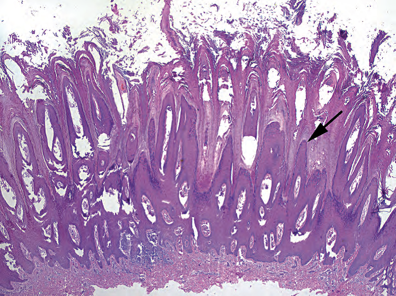
Figure 27.17. Verruca vulgaris. The epidermis in this wart is thrown up into sharply pointed spires, which are topped by hyperkeratosis and parakeratosis.
图27.17 寻常疣。疣的表皮被抛向尖顶,尖顶有角化过度和角化不全。
If you cannot quite tell if a lesion is a seborrheic keratosis or a wart, compromise and call it a verrucous keratosis. Verrucous carcinoma, a deceptively innocuous cancer, is not usually in the differential diagnosis for skin: it is mainly seen on mucosal sites.
如果你不清楚病变是脂溢性角化病还是疣,就折衷一下,称之为疣状角化病。疣状癌,一种貌似温良的癌症,通常不在皮肤的鉴别诊断中:它主要见于粘膜部位。
皮肤附属器肿瘤(Adnexal Tumors)
Adnexal tumors are a large, mystifying, shape-shifting group of lesions encompassing follicular, eccrine, apocrine, and sebaceous lesions. Some of the more readily identifiable tumors are listed here. Most of these are benign, although carcinomas do exist. Of the carcinomas, many are similar to those found in breast or salivary gland (similar embryologic origin), such as adenoid cystic carcinoma, mucoepidermoid carcinoma, and ductal adenocarcinomas.
附属器肿瘤是一组庞大的、神秘的、形状改变的病变组,包括毛囊、小汗腺、大泌腺和皮脂腺的病变。这里列出了一些较容易识别的肿瘤。大多数是良性,但癌确实存在。其中许多癌类似于乳腺或涎腺中的癌(胚胎起源相似),如腺样囊性癌、粘液表皮样癌和导管腺癌。
The eccrine poroma/acrospiroma/hidradenoma group are tumors of the sweat ducts and get different names depending on where in the dermis or epidermis they arise. They are composed of cells that look similar to keratinocytes but that try to form ducts (usually tiny lumens in a sheet of cells). The cells are streamy, pale, and disorganized, not unlike usual ductal hyperplasia in the breast (Figure 27.18).
小汗腺汗孔瘤/肢端汗腺瘤/汗腺瘤组是汗腺导管肿瘤,根据其在真皮或表皮中的位置而有不同的名称。它们由看似角质细胞的细胞组成,试图形成导管(通常是一片细胞中的微小管腔)。这些细胞呈流线型、淡染、无组织结构,不像乳腺导管增生(图27.18)。
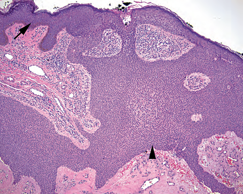
Figure 27.18. Poroma. This eccrine tumor is continuous with the epidermis, which can be seen at left (arrow). The tumor cells (arrowhead) are uniform, small, round, and pale and in some areas may form rudimentary duct spaces.
图27.18 汗孔瘤。这种小汗腺肿瘤与表皮相连续(左侧,箭)。肿瘤细胞(箭头)均匀、小、圆、淡染,在某些区域可能形成残留的导管腔。
Eccrine spiroadenomas are “blue cannonballs in the dermis.” Tumor balls consist of two basaloid cell lineages (often hard to separate) and have noticeable cords and droplets of hyaline basement membrane substance running through them (Figure 27.19). A related lesion is the cylindroma.
小汗腺螺旋腺瘤是“真皮中的蓝色炮弹”。肿瘤球由两个基底细胞谱系组成(通常很难分离),并有明显的透明基底膜物质索状和液滴穿过它们(图27.19)。一个相关的病变是圆柱瘤。
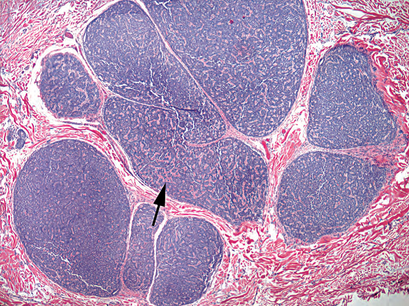
Figure 27.19. Eccrine spiroadenoma. “Cannonballs in the dermis” is the catch phrase for this tumor. Like the poroma, the cells are small and bland. Cords of hyaline pink basement membrane material are seen throughout the tumor (arrow).
图27.19 小汗腺螺旋腺瘤。俗称“真皮中的炮弹”。像汗孔瘤一样,细胞小而温和。整个肿瘤中可见条索状粉红色透明基底膜物质(箭)。
The cylindroma (“jigsaw puzzle”) also has basaloid (blue) nests in the dermis, also with two cell populations and basement membrane matrix. However, the tumor nests are mosaic in shape.
圆柱瘤(“拼图”)也是真皮中的基底样(蓝色)细胞巢,也有两种细胞群和基底膜基质。然而,肿瘤巢的形状是镶嵌的。
The syringoma is a collection of round, dilated tubules in the dermis with characteristic comma-like or tadpole tails (Figure 27.20). Trichoepithelioma is a benign tumor of hair follicle differentiation that looks a lot like a basal cell carcinoma except with horn cysts, hair formation, little abortive follicles, fibrotic stroma, and a lack of clefting. Microcystic adnexal carcinoma (sclerosing sweat duct carcinoma), although rare, is the malignant one you do not want to miss. It looks similar to syringoma, with tubules and cords of bland cells, and also has horn cysts (Figure 27.21). What differentiates it from syringoma is the deep infiltration of the dermis, so dermatopathologists like to see the base of an adnexal lesion.
汗管瘤是一组圆形、扩张的小管,具有特征性逗号样或蝌蚪样尾巴(图27.20)。毛发上皮瘤是一种毛囊分化的良性肿瘤,除了角囊肿、毛发形成、小的流产的毛囊、纤维化间质和缺乏裂隙外,很像基底细胞癌。微囊性附属器癌(硬化性汗管癌)虽然很少见,却是不能错过的恶性肿瘤。它看似汗管瘤,有形态温和的细胞形成的小管和条索,也有角囊肿(图27.21)。它与汗管瘤的区别在于真皮的深层浸润,因此皮肤病理医生喜欢看到皮肤附属器病变的基底部。
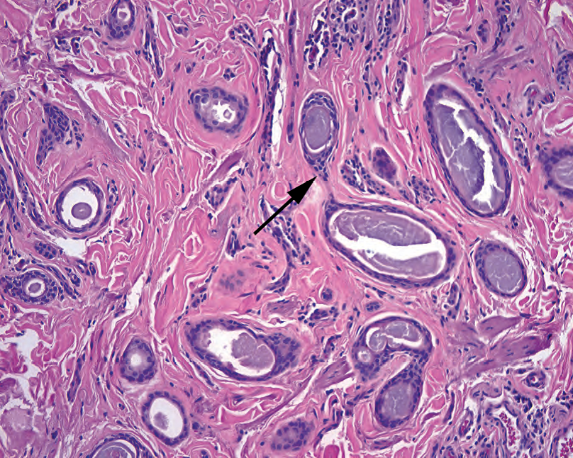
Figure 27.20. Syringoma. Small tubules with comma-like pointed tails within the dermis (arrow).
图27.20 汗管瘤。真皮内具有逗号状尖尾的小管(箭)。
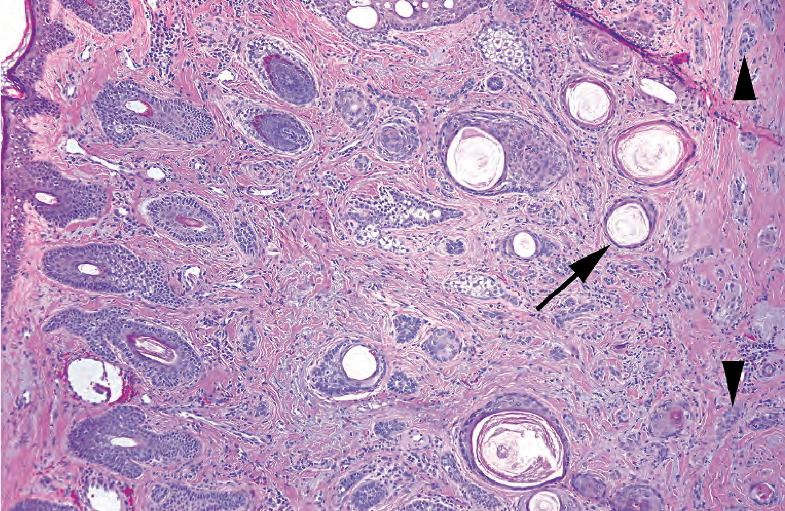
Figure 27.21. Microcystic adnexal carcinoma. A collection of small pale nests of cells can be seen in the dermis, some of them forming horn cysts (arrow). The feature that separates this from a benign lesion is the small nests that are infiltrating deeply into the base of the lesion at right (arrowheads). This small carcinoma infiltrated into the subcutaneous fat (not seen here).
图27.21 微囊性附属器癌。真皮中有一些小的淡染的细胞巢,其中一部分形成角囊肿(箭)。它与良性病变的区别是,图右侧病变基底部有小巢状浸润(箭头)。这个小癌浸润到皮下脂肪(这里看不到)。
囊肿(Cysts)
The most common cysts you will see are the epidermoid cyst (sometimes called epidermal inclusion cyst) and the trichilemmal or pilar cyst (Figure 27.22). The epidermoid cyst is lined by mature squamous epithelium with a granular layer and filled with layers of flaky keratin. 最常见的囊肿是表皮样囊肿(有时称为表皮包含囊肿)、外毛根鞘囊肿(图27.22)。表皮样囊肿衬覆成熟的鳞状上皮,有颗粒层,充满片层状角蛋白。
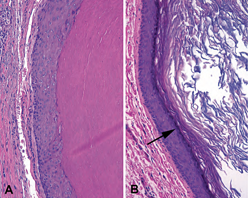
Figure 27.22. Cysts. (A) The trichilemmal cyst has no granular layer, with large pink puffy cells showing an abrupt transition to dense “wet” appearing keratin. (B) The epidermoid cyst more closely resembles epidermis, with a granular layer (arrow) and layers of “dry” flaky keratin.
图27.22 囊肿。(A)外毛根鞘囊肿没有颗粒层,粉红色的肥胖细胞突然转变为致密的“湿的”角蛋白。(B)表皮样囊肿更像表皮,有颗粒层(箭)和“干的”片层状角蛋白。
The pilar cyst is lined by plump pillowy keratinocytes with no granular layer and is filled with dense compact keratin.
皮发囊肿衬覆丰满的枕状角质细胞,无颗粒层,充满致密的角蛋白。
真皮肿瘤(Dermal Tumors)
Probably the three most common benign soft tissue tumors of the dermis are the dermatofibroma, the neurofibroma, and the hemangioma. More information about soft tissue tumors can be found in Chapter 28.
真皮最常见的三种良性软组织肿瘤可能是皮肤纤维瘤、神经纤维瘤和血管瘤。有关软组织肿瘤的更多信息,参见第28章。
Dermatofibromas appear as an ill-defined blue haze in the dermis. On higher power, the blue haze is made up of tiny swarming nondescript cells that infiltrate the collagen and tend to packet it into thick bundles (Figure 27.23). The overlying epidermis may be hyperpigmented and hypertrophic (hence its presentation as a brown nodule). Dermatofibrosarcoma protuberans is the malignant counterpart of this lesion and is more deeply infiltrative, wrapping around the subcutaneous fat in a characteristic pattern (Figure 27.24).
皮肤纤维瘤就像真皮内界限不清的蓝色薄雾。高倍,蓝雾由微小的、成群结队的、难以描述的细胞组成,这些细胞浸润胶原蛋白,并倾向于将其包裹成厚束(图27.23)。上方表皮可能色素沉着且肥大(因此呈棕色结节)。隆突性皮肤纤维肉瘤是该病变的恶性对应物,浸润较深,以特征性模式包裹皮下脂肪(图27.24)。

Figure 27.23. Dermatofibroma. This poorly circumscribed tumor creates a blue haze in the dermis (outlined by arrowheads), and the epidermal rete above it may become pigmented and prominent (arrow). The lesion mainly stops at the subcutaneous fat. Inset: The cells infiltrate between collagen bundles but have small pale round-to-oval nuclei.
图27.23 皮肤纤维瘤。这个边界不清的肿瘤在真皮中形成蓝色薄雾(箭头所示的轮廓),其上方可能有明显的上皮脚伴色素沉着(箭)。病变主要止于皮下脂肪。插图:细胞浸润在胶原束之间,核小,淡染,圆形至椭圆形。
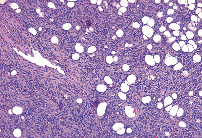
Figure 27.24. Dermatofibrosarcoma protuberans (DFSP). The DFSP is more cellular than the dermatofibroma and shows a prominent storiform pattern of spindled cells infiltrating between fat cells.
图27.24 隆突性皮肤纤维肉瘤(DFSP)。DFSP比皮肤纤维瘤更加细胞丰富,梭形细胞浸润在脂肪细胞之间,形成明显的的网状结构。
Neurofibromas more often appear as a pale or grey nodule in the dermis, more defined than the dermatofibroma. It displaces the dermis rather than infiltrating it. The individual cells have wavy nuclei and wavy collagen, like overstretched elastic (Figure 27.25).
神经纤维瘤通常表现为真皮中的浅染或灰色结节,比皮肤纤维瘤的界限更清楚。它取代真皮而不是浸润真皮。单个细胞有波汶状核和波浪状胶原,就像过度拉伸的弹性纤维(图27.25)。
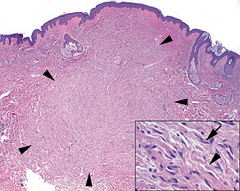
Figure 27.25. Neurofibroma. Like the dermatofibroma, this diffuse neurofibroma is a poorly defined dermal tumor (arrowheads), but, unlike the dermatofibroma, it tends to appear more pale than the surrounding dermis. Inset: On higher power, the tapering, undulating nuclei (arrow) are visible, as is the background of wavy collagen fibers (arrowhead).
图27.25 神经纤维瘤。正如皮肤纤维瘤,这例弥漫性神经纤维瘤是一种界限不清的皮肤肿瘤(箭头所示的轮廓),但与皮肤纤维瘤不同,它往往比周围的真皮更淡染。插图:高倍,核一端逐渐变细、起伏状(箭头),背景中胶原纤维(箭头)也是波浪起伏的。
Hemangiomas are a proliferation of well-formed, dilated capillaries in the dermis (Figure 27.26). There are many variants. The malignant counterpart, angiosarcoma, is more cellular and has anastomosing channels lined by plump cells. Early Kaposi’s sarcoma is so subtle it is basically invisible to the inexperienced and is not likely to simulate a hemangioma (Figure 27.27). Pyogenic granuloma, or lobular capillary hemangioma, is a common benign lesion that may be very cellular and inflamed but is identified as benign due to its rounded and circumscribed periphery.
血管瘤是真皮内的一种毛细血管增殖,这些毛细血管的结构完好,管腔扩张(图27.26)。有许多变异型。对应的恶性肿瘤是血管肉瘤,细胞更多,血管交叉吻合,衬覆丰满的细胞。早期Kaposi肉瘤非常细微,缺乏经验的人可能视而不见,不太像血管瘤(图27.27)。化脓性肉芽肿,或称小叶毛细血管瘤,是一种常见的良性病变,可能是细胞性和炎症性的,但由于其周围呈圆形和局限性而被确定为良性。

Figure 27.26. Capillary hemangioma. There is a collection of discrete, well-formed, dilated capillaries under the epidermis.
图27.26 毛细血管瘤。表皮下有一组离散的、结构完好的、扩张的毛细血管。
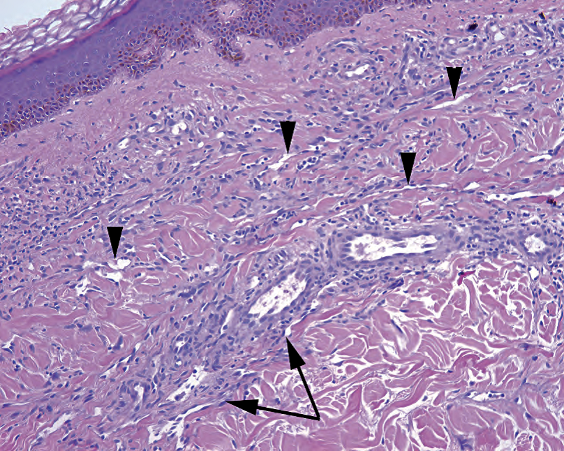
Figure 27.27. Kaposi’s sarcoma. The subtle cellularity to the dermis is actually composed of slit-like vascular spaces with bland endothelium (arrowheads). The slit-like spaces are accentuated around existing capillaries (arrows), the “promontory sign.”
图27.27 卡波西肉瘤。与正常真皮相比,轻微的细胞增多,实际上是由裂隙状血管腔和形态温和的内皮组成(箭头)。裂隙状腔隙在现有的毛细血管周围更明显(箭),即“海角征”
(译注:promontory sign,海角征:卡波西肉瘤中,一个新生血管围绕着一个已经存在的血管腔;换言之,原先存在的血管或附属器结构突入新生血管腔内)
非肿瘤性皮肤病简介:炎症和损伤的模式(A Brief Introduction to Nonneoplastic Skin: Patterns of Inflammation and Injury)
This section is simply a primer on the terminology and classification of the inflammatory skin diseases; the details are beyond the scope of this chapter. These diagnoses are heavily influenced by the clinical presentation, so the goal here is to understand the histologic families of disease. Arriving at a specific diagnosis requires clinical information and, usually, fellowship training.
本节只是炎症性皮肤病的术语和分类的入门;本章不讨论细节。这些诊断受临床表现的严重影响,因此这里的目标是了解疾病的组织学家族。达到一个特定的诊断需要临床信息,通常还需要专科培训。
表皮损伤(Injury to the Epidermis)
The epidermis can become acutely damaged or inflamed. The result is edema, or spongiosis. This is seen as an accentuation of the spaces between keratinocytes. Severe edema can cause intraepidermal vesicles to form (i.e., poison ivy), which have to be distinguished from true bullae (see later).
表皮可能会急性损伤或发炎。结果是水肿或海绵形成(或译作“棘层水肿”)。一般认为这是角质细胞之间空隙变得更明显。严重水肿可能导致表皮内小泡(如,接触毒葛),必须区分真的大疱(见下文)。
If this process continues for a while, the epidermis becomes less edematous and more hyperplastic as it responds to the chronic insult. The hyperplasia is in the form of thickening of the epidermis and elongation of the rete and is called acanthosis, usually accompanied by hyperkeratosis (a protective thickening of the keratin layer).
如果这个过程持续一段时间,表皮水肿会减轻,增生更明显,这是对慢性损伤的反应。这种增生表现为表皮增厚和上皮脚延长,称为棘皮病,通常伴有角化过度(角质层的保护性增厚)。
These changes make up the spectrum of acute to chronic spongiotic dermatitis, clinically eczema, and there is a large differential. Usually the pathologist signs the biopsy out descriptively, and the dermatologist combines that with the clinical features to diagnose it. Psoriasis fits in here, as it has histologic overlap with chronic spongiotic dermatitis, especially when partially treated.
这些变化构成了急性至慢性海绵状皮炎的谱系,临床上表现为湿疹,并有很大的差异。病理医生对活检通常签发描述性报告,皮肤科医生结合活检报告和临床特征进行诊断。例如银屑病,它与慢性海绵状皮炎有组织学重叠,尤其是在部分治疗时。
Some inflammatory processes attack the basal layer of keratinocytes. This pattern is called interface dermatitis. Interface dermatitis has two predominant patterns that may overlap. One is an intense lymphocytic infiltrate at the DEJ, which is called lichenoid inflammation or dermatitis. The second is a vacuolar degeneration of the basal cells, or vacuolar dermatitis. Both of these patterns result in a ragged DEJ, dyskeratotic or necrotic basal cells trapped in the epidermis (colloid bodies), and a cleavage plane along the DEJ if the damage is severe enough. This can be mistaken for a bullous disease, which is a different process.
一些炎症过程攻击角质细胞的基底层。这种模式称为界面皮炎。界面皮炎有两种主要类型,可能重叠。一种是在DEJ处有强烈的淋巴细胞浸润,称为苔藓样炎症或苔藓样皮炎。第二种是基底细胞的空泡变性,或空泡性皮炎。这两种模式都会导致DEJ参差不齐、角化不良或坏死的基底细胞陷入表皮内(胶样小体),如果损伤很严重,则沿着DEJ形成分裂。这可能被误认为大疱性疾病,这是一个不同的过程。
The prototypical lichenoid dermatitis is lichen planus. Vacuolar dermatitis has a wider differential, including acute graft-versus-host disease, lupus, and erythema multiforme.
苔藓样皮炎的原型是扁平苔藓。空泡性皮炎有更广泛的鉴别诊断,包括急性移植物抗宿主病、狼疮和多形性红斑。
A third pattern is the dissolution of the intercellular bridges that link the keratinocytes. The cells break apart and round up into individual cells, a process called acantholysis. This process is often antibody mediated, so immunofluorescence is important. The acantholysis can coalesce into large spaces within the epidermis, or bullae. Different diseases cleave the skin within different planes of the epidermis.
第三种模式是连接角质细胞的细胞间桥的溶解。这些细胞分开,形成单个细胞的聚集,这个过程称为棘层松解。这个过程通常是抗体介导的,因此免疫荧光检查很重要。棘层松解可在表皮内合并成大的空隙,或大疱。不同的疾病会在表皮的不同层面切割皮肤。
This group includes the inflammatory bullous diseases, such as pemphigus, bullous pemphigoid, and dermatitis herpetiformis. A noninflammatory bullous disease is porphyria cutanea tarda. There is also familial acantholytic disease (Hailey-Hailey and Darier diseases), transient acantholytic disease (Grover’s disease), and focal acantholytic lesions (warty dyskeratoma).
这组包括炎性大疱性疾病,如天疱疮、大疱性类天疱疮和疱疹样皮炎。非炎症性大疱性疾病是迟发性皮肤卟啉症。还有家族性棘层溶解病(Hailey-Hailey病和Darier病)、一过性棘层溶解病(Grover病)和局灶性棘层溶解病(疣状角化不良瘤)。
真皮炎症(Inflammation of the Dermis)
The patterns of injury discussed in this section are limited to the dermis, usually with a fairly unremarkable epidermis. Many diseases begin with a nonspecific pattern of perivascular lymphocytic inflammation in the dermis. It is the first sign that the skin is upset. Some diseases never declare themselves beyond this stage, such as polymorphous light eruption and urticaria. If the inflammation progresses to involve neutrophils and actual damage to the vessels, the disease is called a vasculitis. Leukocytoclastic vasculitis, in which the vessels show fibrinoid necrosis and nuclear debris (karyorrhexis), has a wide clinical differential diagnosis.
本节讨论的损伤模式局限于真皮,通常表皮相对不明显。许多疾病始于真皮内血管周围的非特异性淋巴细胞性炎症。这是皮肤不适的第一个迹象。有些疾病在这个阶段之后不再继续,如多形性光疹和荨麻疹。如果炎症进展到涉及中性粒细胞和血管的实际损伤,这种疾病称为血管炎。白细胞裂解性血管炎,其中血管显示纤维素样坏死和核碎屑(核碎裂),具有广泛的临床鉴别诊断。
Inflammatory infiltrates of the dermis are classified based on the type of infiltrate. A dense neutrophilic infiltrate is a neutrophilic dermatosis, such as Sweet’s syndrome. Granulomatous inflammation may indicate infection, foreign body response, sarcoidosis, or granuloma annulare. Mycosis fungoides, or cutaneous T-cell lymphoma, can have numerous appearances, but a dense lymphocytic infiltrate should trigger a workup for mycosis fungoides.
真皮的炎性浸润根据浸润类型进行分类。密集的中性粒细胞浸润是一种中性粒细胞性皮肤病,如Sweet综合征。肉芽肿性炎症可能表提示感染、异物反应、结节病或环状肉芽肿。蕈样肉芽肿,或皮肤T细胞淋巴瘤,可有多种表现,但密集的淋巴细胞浸润应引发蕈样肉芽肿的进一步检查。
Some diseases involve alteration of the collagen of the dermis. These include scar, keloid, scleroderma or morphea, and lichen sclerosus. Chronic graft-versus-host disease can look like scleroderma as well.
有些疾病涉及真皮胶原的改变。这些包括瘢痕、瘢痕疙瘩、硬皮病或局限性硬皮病,以及硬化性苔藓。慢性移植物抗宿主病看起来也像硬皮病。
深部皮下组织炎症(脂膜炎)(Inflammation of the Deep Subcutaneous Tissue (Panniculitis))
Panniculitis is divided into septal, where the inflammation is mostly in the fibrous septae between fat lobules, and lobular, where the fat itself is involved. The classic septal panniculitis is erythema nodosum. Lupus profundus, or deep lupus, is a lobular panniculitis.
脂膜炎分为间隔型和小叶型,前者炎症主要发生在脂肪小叶之间的纤维间隔,后者脂肪本身受累。典型的间隔性脂膜炎是结节性红斑。深部狼疮是一种小叶性脂膜炎。
来源:
The Practice of Surgical Pathology:A Beginner’s Guide to the Diagnostic Process
外科病理学实践:诊断过程的初学者指南
Diana Weedman Molavi, MD, PhD
Sinai Hospital, Baltimore, Maryland
ISBN: 978-0-387-74485-8 e-ISBN: 978-0-387-74486-5
Library of Congress Control Number: 2007932936
© 2008 Springer Science+Business Media, LLC
仅供学习交流,不得用于其他任何途径。如有侵权,请联系删除
本站欢迎原创文章投稿,来稿一经采用稿酬从优,投稿邮箱tougao@ipathology.com.cn
相关阅读
 数据加载中
数据加载中
我要评论

热点导读
-

淋巴瘤诊断中CD30检测那些事(五)
强子 华夏病理2022-06-02 -

【以例学病】肺结节状淋巴组织增生
华夏病理 华夏病理2022-05-31 -

这不是演习-一例穿刺活检的艰难诊断路
强子 华夏病理2022-05-26 -

黏液性血性胸水一例技术处理及诊断经验分享
华夏病理 华夏病理2022-05-25 -

中老年女性,怎么突发喘气困难?低度恶性纤维/肌纤维母细胞性肉瘤一例
华夏病理 华夏病理2022-05-07


共0条评论