外科病理学实践:诊断过程的初学者指南 | 第24章 神经内分泌肿瘤
第24章 神经内分泌肿瘤(Neuroendocrine Neoplasms)
一般定义(General Definitions)
The subject of neuroendocrine neoplasms, starting with the definition of what neuroendocrine means, is thoroughly confusing to the beginner. This chapter reviews the basic concepts and definitions pertaining to this subject.
从神经内分泌的定义开始,神经内分泌肿瘤这个主题对初学者来说是完全混乱的。本章回顾这个主题相关的基本概念和定义。
Let us start with a definition of neuroendocrine. As the term implies, there are two components: “neuro” and “endocrine.” The “endocrine” quality refers to the secretory nature of neuroendocrine cells: they produce and secrete peptides and amines. The “neuro” quality refers to their ultrastructural similarity to neurons: neuroendocrine cells store their secretory products in granules (i.e., dense-core granules), which bear resemblance to synaptic vesicles. Neuroendocrine cells are different from neurons structurally (no processes) and by the fact that the secretory mode is paracrine rather than synaptic. Also note that not all that secretes is neuroendocrine: for example, thyroid and adrenal cortex are not neuroendocrine because their cells do not possess neurosecretory granules (they are simply endocrine). Thus, at the most basic level, neuroendocrine cells are defined as the presence of neurosecretory granules in nonneurons. Tumors derived from these cells have a characteristic “neuroendocrine morphology” and share expression of “neuroendocrine markers.”
让我们从神经内分泌的定义开始。顾名思义,有两个组成部分:“神经”和“内分泌”。内分泌从定性方面说明神经内分泌细胞的分泌性质:它们产生和分泌肽和胺。“神经”是指它们与神经元在超微结构上的相似性:神经内分泌细胞将其分泌产物储存在颗粒(即致密的核心颗粒)中,这些颗粒与突触小泡相似。神经内分泌细胞在结构上不同于神经元(无突起),其分泌方式是旁分泌而不是突触。还要注意的是,并非所有的分泌都是神经内分泌的:例如,甲状腺和肾上腺皮质不是神经内分泌的,因为它们的细胞没有神经分泌颗粒(它们只是内分泌)。因此,在最基本的水平上,神经内分泌细胞被定义为非神经元中存在神经分泌颗粒。来源于这些细胞的肿瘤都有特征性“神经内分泌形态学”,都表达“神经内分泌标记物”。
神经内分泌标记物(Neuroendocrine Markers)
In the past, neurosecretory granules were identified by electron microscopy and special stains. Currently these methods have been completely supplanted by immunohistochemical markers. These are called neuroendocrine markers and they include synaptophysin (SYN), chromogranin (CHR), neural-specific enolase (NSE), and CD56 (SYN and CHR specifically recognize dense-core granules). Note that these markers also recognize true neurons and neuroblastic cells (primitive neurons).
过去,神经分泌颗粒是通过电子显微镜和特殊染色来鉴定的。目前这些方法已被免疫组化标记物完全取代。后者称为神经内分泌标记物,包括突触素(SYN)、嗜铬素(CHR)、神经特异性烯醇化酶(NSE)和CD56(SYN和CHR特异性识别致密核心颗粒)。请注意,这些标记也识别真正的神经元和神经母细胞(原始神经元)。(译注,NSE特异性差,已经很少使用)
神经内分泌形态学(Neuroendocrine Morphology)
Morphologically, unlike adenocarcinoma or squamous cell carcinoma, there is no single feature that defines neuroendocrine neoplasms as a group. Instead, neuroendocrine morphology is defined by a constellation of several cytologic and architectural features:
与腺癌或鳞状细胞癌不同,并没有某个单一的特征可以定义神经内分泌肿瘤的形态学。相反,神经内分泌形态学是由几个细胞学和结构特征的组合共同定义的:
Neuroendocrine cytology
神经内分泌细胞学
Overall nuclear uniformity/monotony with smooth nuclear contours (unlike typical adenocarcinomas or squamous carcinomas)
总体上,核均匀/单一,核轮廓光滑(不同于典型的腺癌或鳞癌)
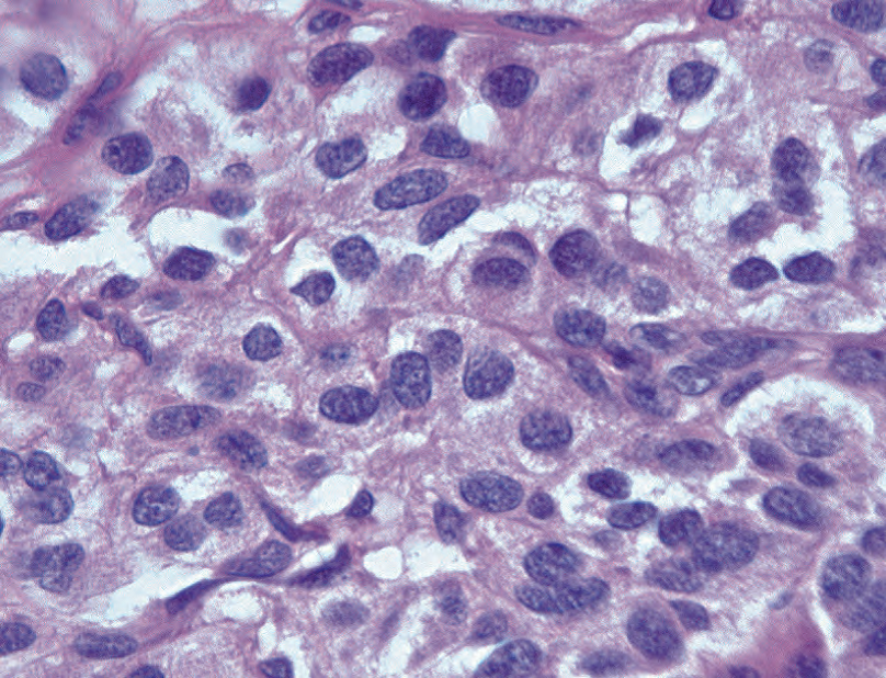
Evenly dispersed, finely speckled “salt and pepper” nuclear chromatin without prominent nucleoli (Figure 24.1)
均匀分散、细腻的点彩状“椒盐样”核染色质,无明显核仁(图24.1)

Figure 24.1. Classic neuroendocrine nuclei, with smooth oval nuclear borders, chromatin that is finely speckled throughout (“salt and pepper”), and no nucleoli.
图24.1 典型的神经内分泌核,有平滑的卵圆形核边界,染色质呈细腻的点彩状(“椒盐样”),没有核仁。
Cytoplasmic granularity (corresponding to “neurosecretory granules”; variably evident)
颗粒性细胞质(对应于“神经分泌颗粒”;不同程度地明显的颗粒性)
Neuroendocrine architecture
神经内分泌结构
Formation of nests, rosettes, and ribbons/trabeculae
形成巢状、菊形团和缎带样/小梁状
Prominent vascularity (in keeping with their secretory nature)
显著的血管(便于分泌物进入血液)
The appearance may be thought of vaguely recapitulating the normal neuroendocrine structures. Recall that nesting is a feature of normal adrenal medulla, and subtle trabecular/ribbonlike structures are present in the islets of Langerhans. The presence of rosettes is not easily explained by resemblance to normal neuroendocrine structures but may relate to their neural lineage, as many neuroglial tumors tend to form rosettes.
这种现象可能认为肿瘤大致再现了正常的神经内分泌结构。回忆一下,正常肾上腺髓质特征之一是成巢,胰岛存在细微的小梁状/缎带状结构。菊形团不像正常神经内分泌结构,但可能与神经谱系有关,因为许多神经胶质肿瘤倾向于形成菊形团。
The morphology features listed apply to both low-grade (carcinoid, pancreatic neuroendocrine neoplasm, pheochromocytoma) and high-grade (small cell neuroendocrine carcinoma, Merkel cell carcinoma) neuroendocrine neoplasms. Whereas the defining neuroendocrine features are usually very obvious in low-grade neoplasms, they may be quite subtle to barely detectable in high-grade neuroendocrine neoplasms. Nevertheless, even when high grade, the overall uniformity and dispersed finely granular chromatin should be preserved, and at least a hint at neuroendocrine architecture (in the form of nesting, rosettes, or ribbons) is usually present.
上述形态学特征适用于低级别(类癌、胰腺神经内分泌肿瘤、嗜铬细胞瘤)和高级别(小细胞神经内分泌癌、默克尔细胞癌)神经内分泌肿瘤。虽然定义性神经内分泌特征在低级别肿瘤中通常非常明显,但在高级别神经内分泌肿瘤中可能非常细微,几乎看不出。然而,即使是高级别,也应保持总体上均质性和分散的细颗粒状染色质,并且通常至少存在神经内分泌结构的迹象(成巢、菊形团或缎带的形式)。
What makes recognition of neuroendocrine neoplasms as a group so challenging is the fact that neuroendocrine morphology may be extremely subtle. For example, some neuroendocrine neoplasms rarely display typical neuroendocrine architecture (e.g., Merkel cell carcinoma typically has no nests, rosettes, or trabeculae). In addition, some neuroendocrine neoplasms frankly violate the basic rules of neuroendocrine morphology (e.g., higher grade neuroendocrine neoplasms, most notoriously large cell neuroendocrine carcinoma of the lung, do have prominent nucleoli). Therefore, diagnosis frequently hinges on recognition of subtle morphologic clues and confirmation with immunostains.
作为一组肿瘤,神经内分泌肿瘤的识别具有挑战性,是因为神经内分泌形态学可能极其微妙。例如,一些神经内分泌肿瘤几乎不显示典型的神经内分泌结构(例如,Merkel细胞癌通常没有巢状、菊形团或小梁状)。此外,一些神经内分泌肿瘤明显违反了神经内分泌形态学的基本规则(例如,高级神经内分泌肿瘤,最著名的肺大细胞神经内分泌癌,确实有显著的核仁)。因此,诊断通常取决于对细微形态学线索的识别和免疫染色的确认。
神经内分泌细胞与肿瘤(Neuroendocrine Cells and Neoplasms)
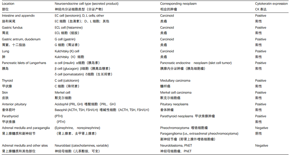
What are the tissues that qualify as neuroendocrine? In addition to neuroendocrine organs (adrenal medulla and paraganglia), the neuroendocrine system includes the so-called diffuse neuroendocrine system. The term diffuse neuroendocrine system refers to neuroendocrine cells dispersed singly or in clusters throughout the body, including pancreas (islets of Langerhans), thyroid (C cells), lungs (Kulchitsky cells), skin (Merkel cells), gastrointestinal tract (many types), and so forth. In fact, neuroendocrine neoplasms may arise in any organ (e.g., prostate, breast, other). The broad definition of neuroendocrine cells also includes parathyroid and anterior pituitary glands. The only endocrine (peptide-secreting) organs excluded as clearly nonneuroendocrine are thyroid gland, adrenal cortex, and the steroid-producing cells of testes and ovaries. Table 24.1 summarizes the major neuroendocrine cell types and their corresponding neoplasms.
什么组织可以定性为神经内分泌?除了神经内分泌器官(肾上腺髓质和副神经节),神经内分泌系统还包括所谓的弥漫神经内分泌系统。这个术语是指分散在全身的单个或成簇的神经内分泌细胞,包括胰腺(胰岛)、甲状腺(C细胞)、肺(Kulchitsky细胞)、皮肤(Merkel细胞)、胃肠道(多种类型)等。事实上,神经内分泌肿瘤可能发生在任何器官(如前列腺、乳腺等)。神经内分泌细胞的广义定义还包括甲状旁腺和垂体前叶。唯一被排除在外的内分泌(肽分泌)器官是甲状腺、肾上腺皮质以及睾丸和卵巢的类固醇生成细胞,显然不是神经内分泌器官。表24.1总结了主要的神经内分泌细胞类型及其相应的肿瘤。
(译注:The normal neuroendocrine (NE) cells in the lung (known as Kulchitsky cells) are present as rare single cells or small cell clusters of 4 to 10 cells (known as neuroepithelial bodies) within bronchial and bronchiolar epithelium.
肺中的正常神经内分泌(NE)细胞称为Kulchitsky细胞,在支气管和细支气管上皮内以罕见的单细胞或4-10个细胞组成的小细胞簇(称为神经上皮体)存在。
Merkel cells are thought to be a type of skin neuroendocrine cell, because they share some features with nerve cells and hormone-making cells. Merkel cells are found mainly at the base of the top layer of the skin (the epidermis). These cells are very close to nerve endings in the skin.
一般认为默克尔细胞是一种皮肤神经内分泌细胞,因为它们与神经细胞和激素生成细胞有一些共同的特征。默克尔细胞主要位于皮肤表层(表皮)的底部。这些细胞非常接近皮肤的神经末梢。)
Table 24.1. Major neuroendocrine cell types and corresponding neoplasms.
表24.1 主要的神经内分泌细胞类型和相应的肿瘤。

ACTH, adrenocorticotropic hormone促肾上腺皮质激素; EC, enterochromaffin肠嗜铬素; ECL, enterochromaffin-like肠嗜铬样; FSH, folliclestimulating hormone促卵泡激素; GH, growth hormone生长激素; LH, luteinizing hormone黄体生成素; PNET, primitive neuroectodermal tumors原始神经外胚层肿瘤; PRL, prolactin催乳素; PTH, parathyroid hormone甲状旁腺激素
(译注:D cell: An enteroendocrine cell that produces somatostatin and is found in the pancreatic islets, stomach, and small intestine. Synonym: delta cell; somatostatin cell. D细胞:一种产生生长抑素的肠内分泌细胞,见于胰岛、胃和小肠。同义词:δ细胞;生长抑素细胞。)
L cell: An enteroendocrine cell that produces glucagon-like peptide-1 and is found in the small intestine. This peptide signals the pancreas to secrete insulin after a meal. L细胞:一种产生胰高血糖素样肽-1的肠内分泌细胞,见于小肠。这种肽向胰腺发出信号,在饭后分泌胰岛素。
一个潜在的令人困惑的问题:神经外胚层肿瘤(A Potentially Confusing Issue: Neuroectodermal Tumors)
What about primitive neuroectodermal neoplasms (primitive neuroectodermal tumors and neuroblastoma)? Do these belong to the family of neuroendocrine neoplasms? These tumors do have neurosecretory-type granules (which are SYN/CHR+), and the neuroblastoma secretes catecholamines, so technically they should qualify as neuroendocrine. However, these neoplasms also appear to possess the ultrastructural features of true primitive neurons (such as neurites). Therefore, together with medulloblastoma, they are classified under a separate heading of primitive neural (neuroblastic/neuroectodermal) tumors. One could think of these as neuroendocrine neoplasms with primitive neuronal phenotype.
原始神经外胚层发生的肿瘤(原始神经外胚层肿瘤和神经母细胞瘤)是什么东西?是否属于神经内分泌肿瘤家族?这些肿瘤确实有神经分泌型颗粒(SYN/CHR+),神经母细胞瘤分泌儿茶酚胺,因此从技术上讲,它们应该符合神经内分泌的条件。然而,这些肿瘤似乎也具有真正的原始神经元(如神经突)的超微结构特征。因此,与髓母细胞瘤一起,它们归入原始神经(神经母细胞/神经外胚层)肿瘤的独立标题。可以认为它们是神经内分泌肿瘤伴具有原始神经元表型。
另一个可能令人困惑的问题:神经内分泌肿瘤中的细胞角蛋白表达(Another Potentially Confusing Issue: Cytokeratin Expression in Neuroendocrine Neoplasms)
Another potentially confusing issue with neuroendocrine neoplasms is their status as neural versus epithelial tissues. In the recent past, all neuroendocrine cells were erroneously thought to be neural (neural crest) derived; this is probably what you learned in medical school. However, it appears that neuroendocrine neoplasms actually fall into two groups: the “truly neural” group (pheochromocytoma/paraganglioma) and the “endoderm-derived/epithelial” group (carcinoid, pancreatic endocrine neoplasm, small cell carcinoma, other). This distinction has practical implications: “neural” neuroendocrine neoplasms are cytokeratin negative, whereas “epithelial” neuroendocrine neoplasms are cytokeratin positive.
神经内分泌肿瘤另一个令人困惑的问题是它们属于神经组织还是上皮组织。不久前,所有的神经内分泌细胞被错误地认为是神经(神经嵴)起源;这可能是你在医学院学到的。然而,神经内分泌肿瘤似乎实际上分为两组:“真正的神经”组(嗜铬细胞瘤/副神经节瘤)和“内皮层起源的/上皮”组(类癌、胰腺内分泌肿瘤、小细胞癌,等)。这种区别具有实际意义:“神经源性”神经内分泌肿瘤呈CK阴性,而“上皮源性”神经内分泌肿瘤呈CK阳性。
还有一些(可能令人困惑的)术语(Some More (Potentially Confusing) Terminology)
To complicate matters further, there are a number of terms that have been applied to neuroendocrine cells over the years. These terms are rarely in routine use today, but they may be encountered in the literature (or on the boards):
更复杂的是,神经内分泌细胞曾经有许多术语。现在已经很少使用,但在文献(或专业组)中可能会遇到:
Amine precursor uptake and decarboxylase (APUD) cells and diffuse neuroendocrine system are the terms for neuroendocrine cells scattered throughout the body (like enterochromaffin-like cells in the stomach). An APUD-oma is another term for carcinoid tumor.
胺前体摄取和脱羧酶(APUD)细胞和弥漫神经内分泌系统是指散布在全身的神经内分泌细胞(如胃中的嗜铬样细胞)。APUD瘤是类癌的另一个术语。
Chromaffin is a term applied to adrenal medulla because of its property to stain brown with chromic salts.
嗜铬是用于肾上腺髓质的术语,因为它用铬盐染成棕色。
Enterochromaffin refers to neuroendocrine cells of the intestine with similar properties, hence “entero” (gut).
肠嗜铬细胞是指肠内具有类似性质的神经内分泌细胞,因此加前缀“肠”。
Enterochromaffin-like refers to histamine-secreting neuroendocrine cells of the gastric fundus.
肠嗜铬样是指胃底分泌组胺的神经内分泌细胞。
Argentaffin and argyrophil refer to the property of neuroendocrine cells to take up silver stains without or with a pretreatment step, respectively. Pretreatment gets more cells to stain. Fontana-Masson is a type of argentaffin stain (it also stains melanin), and Grimelius is a type of argyrophil stain. These stains are of historic interest only, because they have been supplanted by immunostains in practice.
嗜银细胞和亲银细胞分别是指神经内分泌细胞在没有或有预处理步骤的情况下吸收银染的特性。预处理使更多的细胞染色。Fontana Masson是一种嗜银染色(它也染色黑色素),Grimelius(六胺银)也是一种嗜银染色。这些染色只具有历史意义,因为它们在实践中已被免疫染色所取代。
选讲一些神经内分泌肿瘤(Select Neuroendocrine Neoplasms)
Neuroendocrine neoplasms encompass such a heterogeneous group of lesions that it may be difficult to see the common thread among them. This section attempts to highlight the common neuroendocrine qualities as well as organ-specific features of neuroendocrine neoplasms.
神经内分泌肿瘤包括一组异质性病变,因此很难看到它们之间的共同点。本节试图强调神经内分泌肿瘤的共同神经内分泌特征以及器官特异性特征。
Note that neuroendocrine neoplasms (particularly low grade) frequently display random nuclear atypia, that is, neuroendocrine-type atypia or pleomorphism. Nuclei are smudgy and have bizarre shapes. Neuroendocrine atypia is degenerative in nature, probably owing to the slow growth rate of these neoplasms, and has no correlation with malignant potential. Neuroendocrine atypia is particularly prominent in pheochromocytoma and paraganglioma; do not mistake this for a feature of high-grade malignancy.
注意,神经内分泌肿瘤(特别是低级别肿瘤)经常表现为随机的核异型性,即,神经内分泌型异型性或多形性。核污浊(模糊不清),形状怪异。神经内分泌异型性本质上是退化性质,可能是由于这些肿瘤生长缓慢,与恶性潜能无关。神经内分泌异型性在嗜铬细胞瘤和副神经节瘤中尤为明显;不要误认为高级别恶性肿瘤的特征。
Expression of neuroendocrine markers is another defining feature of neuroendocrine neoplasms; expression is usually strong in low-grade lesions and may be weak/focal in high-grade lesions. Note that in the case of small cell carcinoma, diagnosis is based predominantly on morphology: if morphology is classic, expression of neuroendocrine markers is not required for diagnosis. The following discussion of specific neuroendocrine neoplasms emphasizes what constitutes their neuroendocrine qualities.
表达神经内分泌标志物是神经内分泌肿瘤的另一个决定性特征;低级别病变中通常强表达,而高级别病变可能较弱/局限表达。注意,对于小细胞癌,诊断主要基于形态学:如果形态学典型,则神经内分泌标记物的表达不是诊断必需的。以下讨论一些特定的神经内分泌肿瘤,强调它们有哪些神经内分泌特征。
高分化神经内分泌肿瘤(类癌)(Well-Differentiated Neuroendocrine Neoplasm (Carcinoid))
This prototypical neuroendocrine tumor has all of the features listed earlier as the hallmarks of neuroendocrine differentiation, including finely speckled chromatin with no prominent nucleoli, uniform (monotonous) round nuclei with a smooth nuclear membrane, and frequently a plasmacytoid appearance (eccentrically placed nucleus). The architecture may be nests, rosettes, ribbons, or trabeculae. Delicate fibrovascular septae are characteristic. Neuroendocrine markers are usually strongly expressed.
这是典型的神经内分泌肿瘤,具有上述神经内分泌分化的所有特征,包括细腻点彩状染色质,无明显核仁,均匀(单调)圆形核,核膜光滑,常出现浆细胞样外观(核偏心)。结构可以是巢状、菊形团、缎带或小梁。纤细的纤维血管间隔是其特征。神经内分泌标记物通常强烈表达。
低分化神经内分泌癌(小细胞癌)(Poorly Differentiated Neuroendocrine Carcinoma (Small Cell Carcinoma))
Despite the name, diagnosis is not based purely on size, but nuclear size is generally less than three lymphocytes in diameter. The chromatin is finely speckled but is also very dark/ hyperchromatic, which may obscure the “salt and pepper” quality in a surgical specimen.
尽管称为“小”细胞,诊断并不仅仅基于细胞大小,但核大小通常小于三个淋巴细胞的直径。染色质呈细腻的点彩状,但也可能非常黑/深染,这可能会掩盖外科标本中的“椒盐样”染色质。
As in other neuroendocrine neoplasms, there are no prominent nucleoli. The unique features include nuclear molding, high nuclear to cytoplasmic ratio with very scant cytoplasm, numerous mitoses and apoptotic bodies, and frequent crush artifact with DNA streaming (known as the Azzopardi phenomenon). Nests, trabeculae, and rosettes are uncommon. The most reliable immunostain is CD56, but all may be positive. If morphology is classic, expression of neuroendocrine markers is not required for diagnosis.
正如其他神经内分泌肿瘤,小细胞癌没有明显的核仁。其独特的特征包括核镶嵌,高核质比,细胞质非常稀少,大量核分裂和凋亡小体,常见挤压假象(称为Azzopardi现象,挤压造成核内DNA泄露因而深染、流水状)。细胞巢、小梁和菊形团不常见。最可靠的免疫染色是CD56,但可能全部为阳性。如果形态学典型,则神经内分泌标记物的表达不是诊断所必需的。
皮肤默克尔细胞癌(Merkel Cell Carcinoma of Skin)
Merkel cell carcinoma is a small round blue cell tumor. Cytology and architecture overlap with small cell carcinoma. As in small cell carcinoma, nuclear molding, crush artifact, and necrosis are usually present. Cytology is extremely high grade with numerous mitoses and apoptotic bodies (although Merkel cell carcinoma shows less molding than small cell carcinoma). Stains are usually required to distinguish the two.
默克尔细胞癌是一种小圆蓝细胞肿瘤。细胞学和结构与小细胞癌重叠。与小细胞癌一样,通常会出现核镶嵌、挤压假象和坏死。细胞学级别极高,有许多核分裂和凋亡小体(默克尔细胞癌的核镶嵌不如小细胞癌明显)。通常需要免疫染色来区分两者。
Neuroendocrine morphology may be subtle, although neuroendocrine markers are reliably expressed. Hints at the neuroendocrine nature are overall nuclear monotony, despite the high nuclear grade. Although the “salt and pepper” quality of chromatin may be subtle at best, it does show dispersed granularity (so-called dusty look). Also, trabeculae and rosettes may be present (hence the former designation as trabecular carcinoma), although more commonly the pattern is diffuse.
神经内分泌形态学可能是微妙的,但神经内分泌标记物都是可靠表达的。提示神经内分泌本质的线索是总体上呈现核单调性,尽管核级别很高。虽然染色质的“椒盐样”质地最多可能是轻微的,但它确实显示出弥散分布(所谓的粉尘样外观)。此外,可能存在小梁和菊形团(因此以前称为小梁癌),尽管更常见的模式是弥漫性的。
In addition to neuroendocrine markers, Merkel cell carcinoma stains for neurofilament, which is normally a neuronal marker, and CK20 (in classic punctate perinuclear dots). In contrast, small cell carcinoma is negative for neurofilament and CK20 but is positive for TTF-1.
除了神经内分泌标记物,Merkel细胞癌还表达神经丝(NF,通常为神经元标记物)和CK20(典型的核旁点状显色)。相反,小细胞癌不表达NF和CK20,而是TTF-1阳性。
甲状腺髓样癌(Medullary Carcinoma of Thyroid)
Neuroendocrine cytology is as described for carcinoid; speckled chromatin may not be evident in surgical slides due to hyperchromasia but should be more apparent in cytology preparations. Plasmacytoid cytology is common. Neuroendocrine architectural features may be present. The unique features are the presence of amyloid and tendency to form large cellular islands.
它的神经内分泌细胞学正如类癌;手术标本组织学切片由于核深染,点彩状染色质可能不明显,但在细胞学标本中更明显。常见浆细胞样细胞学。可能存在神经内分泌结构特征。独特的特征是存在淀粉样蛋白的存在和倾向于形成大的细胞岛。
大细胞神经内分泌癌(Large Cell Neuroendocrine Carcinoma)
Large cell neuroendocrine carcinoma is one of the neuroendocrine neoplasms that you would not guess had a neuroendocrine nature based on cytology (nuclei are vesicular, not salt and pepper, and have a single prominent nucleolus). Neuroendocrine classification is based on a subtle hint of neuroendocrine architecture (rosettes and nuclear palisading), expression of neuroendocrine markers, and ultrastructural demonstration of dense-core granules.
这种神经内分泌肿瘤,根据细胞学检查,你猜不到它是神经内分泌性质(空泡状核,而不是椒盐样,并有单个明显核仁)。神经内分泌分类基于神经内分泌结构的细微迹象(菊形团和核栅栏状排列)、表达神经内分泌标记物和超微结构显示致密核心颗粒。
嗜铬细胞瘤(肾上腺)/副神经节瘤(肾上腺外)(Pheochromocytoma (Adrenal)/Paraganglioma (Extraadrenal))
Classic neuroendocrine cytology may be barely discernible in a pheochromocytoma. Some nuclei are carcinoid-like in that they are uniform, round, and finely speckled, but many nuclei are more neuron-like by virtue of large size and single prominent nucleolus. The cytoplasm is abundant, granular, and “amphophilic” (lavender). Nuclear pseudoinclusions (cytoplasmic invaginations) are present in 30% of cases. Hyaline globules are common and, if present, distinguish pheochromocytomas from other adrenal neoplasms. Random nuclear atypia is common.
经典的神经内分泌细胞学在嗜铬细胞瘤中几乎不可见。有些核呈类癌样,呈均匀、圆形、细腻的点彩状,但许多核更像神经元,核大,有单个明显的核仁。胞质丰富,呈颗粒状,双染性(淡紫色)。30%的病例出现核内假包涵体(细胞质内陷)。常见透明小球,如有,可区分其他肾上腺肿瘤区分。常见随机的核异型性。
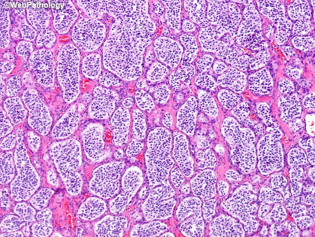
Although paragangliomas are equated with extraadrenal pheochromocytomas, the morphology is not identical. In paraganglioma, the nuclei are much more carcinoid-like. The architecture is nested and occasionally trabecular, but there are no rosettes. The nest pattern is referred to as zellballen, which in German literally means cell balls. The zellballen pattern is highlighted by S100 stain, which reacts with sustentacular/supportive cells outlining the nests (sustentacular cells are not visible on H&E stain).
虽然副神经节瘤等同于肾上腺外嗜铬细胞瘤,但形态并不完全相同。副神经节瘤的细胞核更像类癌。结构成巢,偶有小梁,但没有菊形团。成巢模式称为zellballen,德语意思是细胞球。S100染色显示支持细胞,勾画出成巢模式。HE染色看不出支持细胞。
(译注:Zellballen: Nest-like clusters of uniform, round-to-polygonal chief cells surrounded by delicate richly vascular tissue and sustentacular cells, a pattern characteristic of paraganglioma. 由均匀一致的、圆形到多边形的主细胞组成巢状细胞簇,周围环绕着纤细的富于血管的组织和支持细胞,这是副神经节瘤的特征。补充图片如下)

Marker expression is identical in both tumors. Neuroendocrine markers are positive in the nests (chromaffin cells), and S100 highlights sustentacular cells. Unlike all other neuroendocrine neoplasms (e.g., carcinoid), these tumors are cytokeratin negative.
标记物在两种肿瘤中的表达相同。细胞巢(嗜铬细胞)表达神经内分泌标记物,支持细胞表达S100。不同于所有其他神经内分泌肿瘤(如类癌),这些肿瘤呈CK阴性。
其他类型癌的神经内分泌分化(Neuroendocrine Differentiation in Other Types of Carcinoma)
As discussed earlier, neuroendocrine neoplasms are diagnosed based on (1) morphology and (2) expression of neuroendocrine markers (electron microscopy and special stains are basically obsolete). If morphology and markers are concordant (which is usually the case), one can comfortably diagnose a neuroendocrine neoplasm. However, there are cases in which morphology and neuroendocrine marker expression are discordant. These distressing situations come in two varieties.
如前所述,神经内分泌肿瘤的诊断基于(1)形态学和(2)神经内分泌标记物的表达(电子显微镜和特殊染色基本上已经过时)。如果形态学和标记物一致(通常如此),就可以轻松诊断神经内分泌肿瘤。然而,在某些情况下,形态学和神经内分泌标记物的表达是不一致的。令人痛苦的情况有两种。
First, expression of neuroendocrine markers may be detected incidentally in an otherwise entirely nonneuroendocrine neoplasm (morphologically), such as a classic adenocarcinoma. In most organs, this is thought to represent a type of occult differentiation with no clear c linical significance. These lesions may be signed out as Carcinoma with neuroendocrine differentiation by immunohistochemistry.
首先,完全没有神经内分泌肿瘤的形态学,偶然检测到神经内分泌标记物的表达,例如典型的腺癌。在大多数器官中,视为无明确临床意义的隐匿性分化。可以报告为“癌伴神经内分泌分化(免疫组织化学检测)"。
Conversely, some high-grade neuroendocrine neoplasms, particularly small cell carcinoma, may express neuroendocrine markers only focally or not at all. Nevertheless, classic morphology trumps the lack of marker expression, once other small cell neoplasms have been excluded.
相反,一些高级神经内分泌肿瘤,特别是小细胞癌,可能只是局部表达或根本不表达神经内分泌标记物。然而,一旦排除了其他小细胞肿瘤,经典形态学胜过标记物表达缺失。(译注:诊断原则:形态学和免疫组化不一致时,以形态学为主)
Note that the first scenario is different from finding small cell carcinoma as a component of another type of carcinoma, such as adenocarcinoma or squamous cell carcinoma. The latter situation is considered to be a form of dedifferentiation to a more primitive phenotype, which does carry a worse prognosis and a need for specific therapy. Such cases are signed out as Mixed adenocarcinoma/small cell carcinoma or Adenocarcinoma with small cell component.
注意,第一种情况不同于发现小细胞癌是另一种癌症的组成部分,如腺癌或鳞状细胞癌中发现小细胞癌。可以报告为“混合腺癌/小细胞癌,或腺癌伴小细胞成分”。第二种情况视为一种去分化形式,变成更原始的表型,预后更差,需要特殊治疗。
各种器官中神经内分泌肿瘤的报告术语(Sign-Out Terminology for Neuroendocrine Neoplasms in Various Organs)
A practical issue of note is that clinical behavior of low-grade neuroendocrine neoplasms (carcinoid, pancreatic endocrine neoplasm, pheochromocytoma) is notoriously difficult to predict based on histologic parameters. Generally, the only definitive sign of malignancy is the presence of metastases. This is why neuroendocrine neoplasms are not staged. However, there are a number of site-specific histologic features that are loosely predictive of a more aggressive phenotype. Given the uncertainty of clinical behavior, there is a range of sign-out terminology applied to neuroendocrine neoplasms (which can vary among institutions):
注意的一个实际问题,根据组织学参数,难以预测低级别神经内分泌肿瘤(类癌、胰腺内分泌肿瘤、嗜铬细胞瘤)的临床行为。一般来说,恶性肿瘤唯一明确的标志是存在转移。这就是为什么神经内分泌肿瘤没有分期。然而,有许多特定部位的组织学特征可以粗略地预测更具侵袭性的表型。鉴于临床行为的不确定性,有许多报告术语适用于神经内分泌肿瘤(各机构之间可能有所不同),举例如下:
Primary lung neuroendocrine neoplasms may be signed out as the following:
原发性肺神经内分泌肿瘤可按以下方式签发报告:
Carcinoid tumor (low-grade neuroendocrine carcinoma): Typical neuroendocrine morphology is apparent, with<2 mitoses/10 high-power fields [hpf] and no necrosis.
类癌(低级别神经内分泌癌):典型的神经内分泌形态明显,核分裂<2/10HPF,无坏死。
Atypical carcinoid tumor (intermediate-grade neuroendocrine carcinoma): From 2 to 10 mitoses/10 hpf and/or focal necrosis are present.
非典型类癌(中级别神经内分泌癌):核分裂2-10/10HPF和/或局灶性坏死。
Small cell (high-grade neuroendocrine) carcinoma: More than 10 mitoses/10 hpf, extensive necrosis, and specific small cell features are present.
小细胞癌(高级神经内分泌癌):核分裂>10/10HPF,广泛坏死和特定的小细胞特征。
Large cell (high-grade neuroendocrine) carcinoma: More than 10 mitoses/10 hpf, extensive necrosis, and specific features are present.
大细胞癌(高级神经内分泌癌):核分裂>10/10HPF,广泛坏死,并具有特定特征。
Primary gastrointestinal neuroendocrine neoplasms may be signed out as the following:
原发性胃肠道神经内分泌肿瘤可按以下方式签发:
Carcinoid tumor: This indicates typical neuroendocrine morphology with no features of concern (e.g., size >1–2 cm, vascular invasion, necrosis, etc.).
类癌:有典型的神经内分泌形态,没有值得关注的特征(例如,大小>1–2厘米,血管浸润,坏死,等)。
Carcinoid tumor, Malignant carcinoid tumor, or Well-differentiated neuroendocrine carcinoma: Morphology is of typical carcinoid, but the tumor is behaving badly, such as already metastatic. The sign-out terminology is a contentious issue here (a matter of style). Some sign out a lesion as carcinoid irrespective of how malignant it behaves. Others advocate that if metastases are present, the lesion should be signed out as malignant c arcinoid or well-differentiated neuroendocrine carcinoma. The third opinion is that the latter two categories should be used if any features of concern are present.
类癌、恶性类癌或高分化神经内分泌癌:形态为典型的类癌,但肿瘤行为不好,例如已经转移。在这里,报告术语是有争议的(个人风格)。有人签发类癌,不管其恶性程度如何。其他人认为,如果存在转移,病变应签发恶性类癌或高分化神经内分泌癌。第三种意见是,如果存在任何令人关注的特征,则应使用后两类。
Poorly differentiated neuroendocrine carcinoma: The carcinoma morphologically is frankly high grade. Small cell and large cell neuroendocrine carcinomas are included here.
低分化神经内分泌癌:形态学明显高级别。这里包括小细胞和大细胞神经内分泌癌。
Primary pancreatic neuroendocrine neoplasms may be signed out as the following:
原发性胰腺神经内分泌肿瘤可按以下方式签发:
Well-differentiated pancreatic endocrine neoplasm (islet cell tumor): Typical neuroendocrine morphology is apparent, with no features of concern.
高分化胰腺内分泌肿瘤(胰岛细胞瘤):有明显的典型的神经内分泌形态,无令人关注的特征。
Well-differentiated malignant pancreatic endocrine neoplasm (malignant islet cell tumor): This designation is used if any one of the three definitively malignant features are present: (1) metastases, (2) large vessel vascular invasion, and/or (3) invasion of adjacent organs. Lymphatic invasion, large size, microscopically invasive border, and so forth are suspicions but not diagnostic of malignancy.
高分化恶性胰腺内分泌肿瘤(恶性胰岛细胞瘤):如果存在三种明确恶性特征之一:(1)转移,(2)大血管侵犯,和/或(3)邻近器官侵犯,则使用该名称。淋巴管浸润、肿瘤体积大、显微镜下浸润性边界等都属于可疑恶性,但不是恶性肿瘤的诊断标准。
Poorly differentiated malignant pancreatic endocrine neoplasm (malignant islet cell tumor): morphologically a frankly high-grade carcinoma is present. Small cell and large cell neuroendocrine carcinoma are included here.
低分化恶性胰腺内分泌肿瘤(恶性胰岛细胞瘤):形态学高级别。这里包括小细胞和大细胞神经内分泌癌。
Note that pancreas and intestine do not have an “atypical” category corresponding to the “atypical carcinoid” category in the lung. Nevertheless, some pathologists do flag intestinal and pancreatic lesions as “atypical” based on the lung criteria (discussed earlier).
注意,胰腺和肠没有对应于肺中“非典型类癌”类别的“非典型”类别。然而,一些病理学家确实根据肺部标准(前面讨论过)将肠道和胰腺病变签发为“非典型”。
The issue of metastatic neuroendocrine neoplasm of unknown origin usually arises for hepatic neuroendocrine neoplasms. The differential diagnosis includes intestinal versus pancreatic metastases (primary hepatic neuroendocrine neoplasms are vanishingly rare). Intestinal carcinoids and pancreatic neuroendocrine neoplasms are histologically identical, and hormone expression is generally not reliable. Therefore clinical correlation is required. These lesions are signed out as “metastatic (well or poorly) differentiated neuroendocrine neoplasm (carcinoid vs. pancreatic endocrine carcinoma)”.
通常在肝神经内分泌肿瘤中遇到来源不明的转移性神经内分泌肿瘤的问题。鉴别诊断包括肠转移或胰腺转移(原发性肝神经内分泌肿瘤非常罕见)。肠类癌和胰腺神经内分泌肿瘤在组织学上相同,激素表达通常不可靠。因此,需要结合临床。这些病变签发为“转移性(高分化或低分化)神经内分泌肿瘤(类癌或胰腺内分泌癌)”。
One contentious issue is the use of the term neoplasm versus carcinoma as it pertains to neuroendocrine tumors that are displaying overtly malignant behavior. In principle, these tumors should be called carcinomas (i.e., malignant epithelial neoplasms). This terminology is indeed advocated by some authorities. Some authorities even advocate use of the term lowgrade neuroendocrine carcinoma in place of carcinoid tumors to reflect the potential of any of these tumors to metastasize. In contrast, other authorities stress that the term neoplasm is preferable (regardless of clinical behavior or morphology) in order to draw a clear distinction between these tumors and typical carcinomas, such as pancreatic adenocarcinoma, which behave in a much more aggressive fashion than any neuroendocrine tumor.
一个有争议的问题是使用术语“肿瘤”与“癌”,因为它涉及表现出明显恶性行为的神经内分泌肿瘤。原则上,这些肿瘤应称为癌(即恶性上皮性肿瘤)。这一术语确实是一些学者提倡的。一些学者甚至主张用低级别神经内分泌癌来代替类癌,以反映这些肿瘤转移的可能性。与此相反,其他学者强调肿瘤一词更可取(无论临床行为或形态如何),以便明确区分这些肿瘤和典型癌症,如胰腺癌,后者的行为方式比任何神经内分泌肿瘤都更具侵袭性。
来源:
The Practice of Surgical Pathology:A Beginner’s Guide to the Diagnostic Process
外科病理学实践:诊断过程的初学者指南
Diana Weedman Molavi, MD, PhD
Sinai Hospital, Baltimore, Maryland
ISBN: 978-0-387-74485-8 e-ISBN: 978-0-387-74486-5
Library of Congress Control Number: 2007932936
© 2008 Springer Science+Business Media, LLC
仅供学习交流,不得用于其他任何途径。如有侵权,请联系删除。
本站欢迎原创文章投稿,来稿一经采用稿酬从优,投稿邮箱tougao@ipathology.com.cn
相关阅读
 数据加载中
数据加载中
我要评论

热点导读
-

淋巴瘤诊断中CD30检测那些事(五)
强子 华夏病理2022-06-02 -

【以例学病】肺结节状淋巴组织增生
华夏病理 华夏病理2022-05-31 -

这不是演习-一例穿刺活检的艰难诊断路
强子 华夏病理2022-05-26 -

黏液性血性胸水一例技术处理及诊断经验分享
华夏病理 华夏病理2022-05-25 -

中老年女性,怎么突发喘气困难?低度恶性纤维/肌纤维母细胞性肉瘤一例
华夏病理 华夏病理2022-05-07


共0条评论