外科病理学实践:诊断过程的初学者指南 | 第19章 乳腺(Breast)
第19章 乳腺(Breast)
Breast biopsy specimens come in several sizes. There is the initial core biopsy, which is a large-bore needle biopsy, and the excisional biopsy, which is like a small lumpectomy. Some institutions perform cytologic studies (fine-needle aspirations), but their usefulness is limited, as many breast diagnoses are more architectural than cytologic. Biopsies are performed, with few exceptions, to rule out malignancy; there are almost no other disease processes that require tissue monitoring. A biopsy specimen with carcinoma will trigger either a lumpectomy, in which a portion of the breast is removed (a partial mastectomy, breast-conserving therapy), or a mastectomy. The mastectomy itself may include sentinel lymph nodes or, if the sentinel node is positive, an entire axillary dissection. Biopsy and most lumpectomy specimens are entirely submitted, and anything that is oriented is inked with four to six colors so we can identify all of the margins later. Mastectomies have only two margins, deep and superficial, and are representatively sampled by quadrant.
乳腺活检标本有多种尺寸。最初是粗针穿刺活检,这是用带有针芯的大口径穿刺针进行的穿刺活检,而切除活检则类似于小规模的肿块切除术。一些医疗机构进行细胞学研究(细针穿刺),但它们的用处有限,因为许多乳腺诊断更需要结构而不是细胞学。除了少数例外,需要进行活检以排除恶性肿瘤;除恶性肿瘤外几乎没有其他疾病需要进行组织学监测。活检标本有癌,将触发肿块切除术,它会切除部分乳房(部分乳房切除术,用于保乳治疗),或乳房切除术。乳房切除术本身可能包括前哨淋巴结,如果前哨淋巴结阳性,则进一步进行整个腋窝淋巴结清扫。活检和大部分肿块切除标本都是完整送检的,任何定向的组织部位都会涂抹四到六种颜色墨水,这样我们以后就可以识别所有的切缘。乳房切除术只有两个切缘(深切缘和浅切缘),并按四个象限切取代表性性组织。
正常组织学(Normal Histology)
The breast is sort of a giant specialized sweat gland, and so it has secretory glands (acini or lobules), arranged like grapes, and ducts, like the grape stems. A single bunch of grapes is a terminal duct lobular unit (TDLU; Figure 19.1). The ducts from these TDLUs all converge on the nipple, which has multiple large ducts and smooth muscle for ejecting the milk. The breast of a child or man will have ducts but no lobules. During lactation, the lobules fill up with fatty vacuoles of milk, giving them a very characteristic look usually called secretory or lactational change.
乳房本质上是巨大的特化汗腺,因此它有分泌腺(腺泡或小叶,排列像葡萄簇)和导管(像葡萄茎)。一束葡萄就是一个终末导管小叶单位(TDLU;图19.1)。来自这些TDLU的导管都会聚到乳头,乳头有多个大导管和平滑肌,用于排出奶汁。儿童或男性的乳房只有导管,没有小叶。在哺乳期,乳汁中的脂肪空泡填满小叶,使其呈现非常独特的外观,通常称为分泌改变或泌乳改变。
Each lobule and duct is composed of two cell types, the outer myoepithelial layer and the inner epithelial cells (see Figure 19.1). This is an important feature that can separate an in situ lesion (two cell types) from an invasive one (one cell type). The whole structure is bounded by a basement membrane, which is the boundary between in situ and invasive cancers. While there are unusual myoepithelial tumors, in this chapter we will only cover epithelial lesions.
每个小叶和导管由两种细胞组成,即外层的肌上皮层和内层的上皮细胞(见图19.1)。这个重要特征可以区分原位病变(有两种细胞)和浸润性病变(只有一种细胞)。小叶和导管都以基底膜为界,基底膜是原位癌和浸润性癌的边界。虽然存在少见的肌上皮肿瘤,但在本章中只讨论上皮病变。
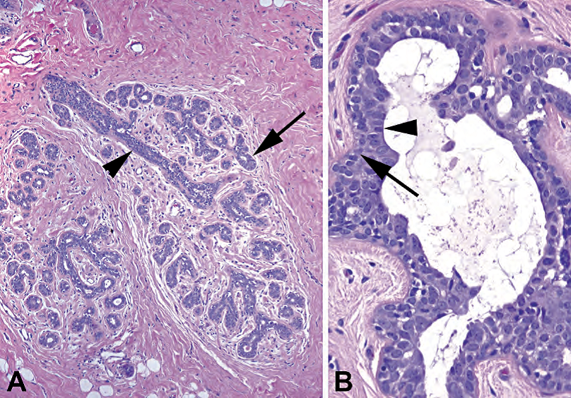
Figure 19.1. Normal breast. (A) The terminal duct lobular unit (TDLU) is arranged like a cluster of grapes, with the duct (arrowhead) as the stem and secretory lobules (arrow) as the grapes. The rounded and circumscribed border of the TDLU is a key feature of noninvasive lesions. (B) The benign breast always has two cell layers, the outer myoepithelial cells (arrow) and the inner epithelial cells (arrowhead). In situ lesions also have two cell layers.
图19.1 正常乳房。(A)终末导管小叶单元(TDLU)的排列像葡萄簇,导管(箭头)就像茎,分泌小叶(箭)作为葡萄。TDLU呈圆形,边界清楚,这是非浸润性性病变的关键特征。(B)良性乳腺病变通常有两层细胞,外层为肌上皮细胞(箭),内层为上皮细胞(箭头)。原位病变也有两层细胞。
活检标本的观察方法(Approach to the Biopsy Specimen)
When signing out a core biopsy, there are certain things that should be included in the diagnosis. For malignant lesions, in situ or invasive, first, it is helpful to give an indication of how differentiated the tumor is. Some institutions do not Elston grade (see later) a core, but you should at least note the nuclear grade (for ductal carcinoma in situ [DCIS]) or whether it is well/moderate/poorly differentiated (for invasive cancer). These three tiers of differentiation correspond roughly to the three Elston grades.
当签发粗针穿刺活检时,诊断中必需包括某些内容。首先,对于恶性病变,无论原位或浸润性,注明肿瘤的分化程度对临床是有帮助的。一些医疗机构不对粗针标本进行Elston分级(见下文),但你至少应该注明核级别(对于导管原位癌,DCIS),或注明高/中/低分化(对于浸润性癌)。高/中/低分化大致对应于Elston分级的三个级别。
Second, if microcalcifications were seen on mammography, you must note whether they are present in the specimen and in what context (such as, “in association with usual duct hyperplasia”). Failure to find the microcalcifications leads to x-raying the block, calling the radiologist, and so forth. Microcalcifications usually are gritty and dark purple, like calcification in other tissues, but occasionally take the sneaky form of calcium oxalate, clear refractile crystals best seen with polarized light (or flipping the condenser down; Figure 19.2).
其次,如果乳房X线检查中发现微钙化,则必须注明标本中是否出现微钙化,以及出现在何种背景下(例如,“普通型导管增生伴微钙化”)。如果切片中找不到微钙化,就需要对时蜡块进行X线检查,电话联系放射科医生,等等。微钙化通常呈砂砾状和深紫色,与其他组织中的钙化相似,但偶见草酸钙型隐匿性钙化,即透明的折光性晶体,最好用偏振光观察(或向下翻转聚光器;图19.2)。
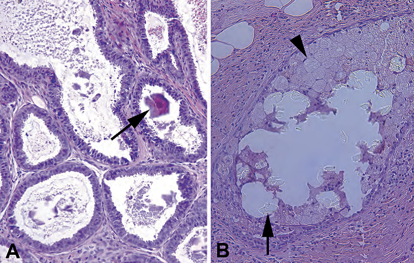
Figure 19.2. Calcifications. (A) Microcalcifications in this columnar cell lesion appear as tiny purple rocks (arrow), which may shatter and drag through the tissue, creating telltale scratches in the H&E stain. (B) Calcium oxalate does not pick up hematoxylin and therefore is only visible with a polarizer or when the condenser is flipped down, as in this photograph. The oxalate crystals (arrow) are seen in a duct space, surrounded by foamy macrophages (arrowhead).
图19.2 钙化。(A)柱状细胞病变中的微钙化,表现为紫色的小石块(箭),可能会粉碎并拖拽组织,在HE染色切片中形成明显的划痕。(B)草酸钙不染苏木素,因此只有在使用偏光镜或将聚光器向下翻转时才可见,如图所示。草酸晶体(箭)可见于导管腔,周围有泡沫状巨噬细胞(箭头)。
Finally, your goal should be to identify the mass or radiographic abnormality the clinicians have detected. If there is no malignancy, you should be looking for some explanation for their findings. Aside from microcalcifications, which do explain a mammographic lesion, you should be looking for anything that could cause a palpable mass, such as fibrosis, cysts or cyst wall, fat necrosis, and benign tumors such as fibroadenomas.
最后,你的目标应该是识别临床医生检测到的肿块或影像学异常。如果没有恶性肿瘤,你应该找到其他病变能够解释其发现。除了微钙化(这确实可以解释乳房X线检出的病变)之外,你应该寻找任何可能引起可触及肿块的东西,如纤维化、囊肿或囊壁、脂肪坏死和良性肿瘤,如纤维腺瘤。
纤维囊性变(Fibrocystic Changes)
Fibrocystic changes are very common in young women, and many palpable lumps turn out to be nothing more than fibrocystic change. These are usually signed out as “Benign breast tissue with fibrocystic changes, including…” and then a list of the features. These features include the following:
纤维囊性变在年轻女性中非常常见,许多可触及的肿块原来只是纤维囊性变,而没有其他任何病变。这些标本通常签发为“良性乳腺组织伴纤维囊性变,包括……”,然后列举一系列特征。这些功能包括:
Fibrosis: Fibrosis consists of dense pink collagen among the lobules.
纤维化:由小叶间致密的粉红色胶原组成。
Cysts: Cysts are often visible grossly, thin walled, and full of clear fluid (Figure 19.3).
囊肿:囊肿通常肉眼可见,壁薄,充满透明液体(图19.3)。
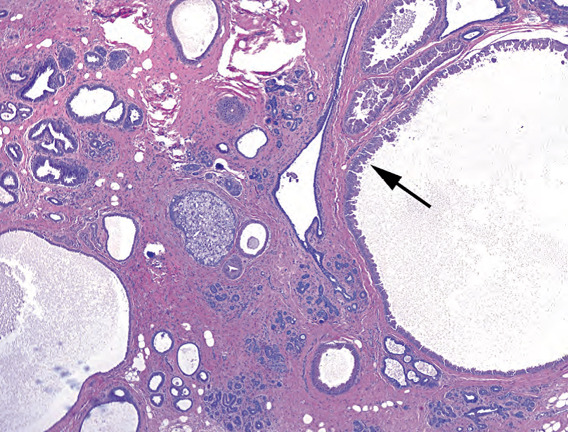
Figure 19.3. Fibrocystic disease. In this example, large dilated duct spaces are visible, some with a lining of apocrine metaplasia (arrow). The stroma is dense and fibrotic (pink).
图19.3 纤维囊性疾病。在本例中,可见巨大的扩张的导管腔,部分导管腔被覆大汗腺化生的上皮(箭)。间质致密,纤维化(粉红色)。
Usual duct hyperplasia: Usual duct hyperplasia is described in detail in a later section.
普通型导管增生:详见下文。
Adenosis (too many glands or lobules) or sclerosing adenosis: Adenosis is a big pitfall because the lobules can look very crowded and worrisome. This is especially true of sclerosing adenosis, in which the proliferative lobules are squeezed together by fibrosis, making them look small and infiltrative (Figure 19.4). The reassuring myoepithelial cell layer can be hard to see. However, sclerosing adenosis should have an overall lobular (circumscribed and rounded) architecture, and myoepithelial cells should be visible in some glands.
腺病(腺体或小叶过多)或硬化性腺病:腺病是一个大陷阱,因为小叶看起来非常拥挤,令人担忧。硬化性腺病尤其如此,增殖的小叶被纤维化挤压在一起,使它们貌似浸润的小管(图19.4)。令人安心的肌上皮细胞层可能很难看到。然而,硬化性腺病整体上应该具有小叶(边界清楚,圆形)结构,并且一些腺体中可见肌上皮细胞。
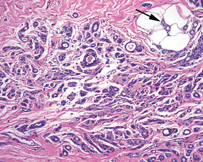
Figure 19.4. Sclerosing adenosis. On high power, this benign lesion looks infiltrative. Tiny tubules are entrapped in a fibrotic stroma, and some tubules are even seen among fat (arrow). Because of the compression, myoepithelial cells are not visible. Clues to the diagnosis include a circumscribed lesion at low power, the lack of desmoplastic (edema and fibrosis) reaction, and an intact myoepithelial cell layer seen on immunostains.
图19.4 硬化性腺病。高倍镜下,这种良性病变貌似浸润性。小管埋陷在纤维化间质中,一些小管甚至见于脂肪中(箭)。由于被挤压,肌上皮细胞不可见。诊断线索包括低倍镜下边界清楚的病变、缺乏促结缔组织增生(水肿和纤维化)反应,以及免疫染色显示的完整肌上皮细胞层。
Apocrine metaplasia: Breasts are just big sweat glands, remember? Apocrine metaplasia means the epithelial cells lining the ducts look like apocrine glands (Figure 19.5); they acquire a lot of bright pink cytoplasm, can get a hobnail profile protruding into the lumen, and have enlarged nuclei with prominent nucleoli (not unlike Hurthle cell change in the thyroid). It is important to recognize this entity as a metaplastic, not a dysplastic, change.
大汗腺化生:乳房只是一个巨大的汗腺,还记得吗?大汗腺化生是指导管上皮细胞看似大汗腺(图19.5);它们获得大量的亮粉色的细胞质,可以形成突向导管腔的鞋钉状轮廓,并且核增大,核仁显著(就像甲状腺的嗜酸细胞改变)。重要的是识别其化生性本质,而不是异型增生改变。
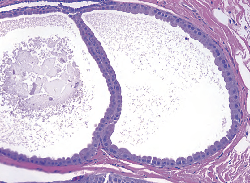
Figure 19.5. Apocrine metaplasia in fibrocystic disease. The epithelial cells lining the dilated duct are large and plump, with abundant dark pink cytoplasm, and round nuclei with prominent nucleoli. Secretions (the granular schmutz in the lumen) are common.
图19.5 纤维囊性疾病中的大汗腺化生。扩张导管被覆的上皮细胞大而丰满,胞质丰富,深粉红色,核圆,核仁显著。常见分泌物(导管腔中的颗粒状污物)。
Fibroadenomas: A fibroadenoma is a biphasic (two cell types) proliferative lesion. The ducts are proliferating (-adenoma), as is the stroma (fibro-). (A similar lesion in the ovary is called an adenofibroma.) This benign tumor has thin, branching ducts set in a sparsely cellular fluffy pink stroma (Figure 19.6). The ducts often have a myxoid pale halo around them, and the proliferative stroma compresses the ducts into slits. Old fibroadenomas may become hyalinized and calcified. Fibroadenomas can occur alone or in association with fibrocystic changes.
纤维腺瘤:纤维腺瘤是一种双相(两种细胞类型)增殖性病变。导管增殖(故称为“腺瘤”),间质也增殖(故加上前缀“纤维”)。(卵巢中类似的病变称为腺纤维瘤。)这种良性肿瘤镜下表现为细长的分支导管位于细胞稀疏的疏松的粉红色间质中(图19.6)。导管周围常有粘液样淡染间质(译注:这是特化的小叶内间质),间质增殖将导管挤压成缝隙状。陈旧性纤维腺瘤的间质可能变成纤维化和胶原化、玻璃样变和钙化(译注,此句有补充)。纤维腺瘤可以单独发生,也可以伴随纤维囊性改变。
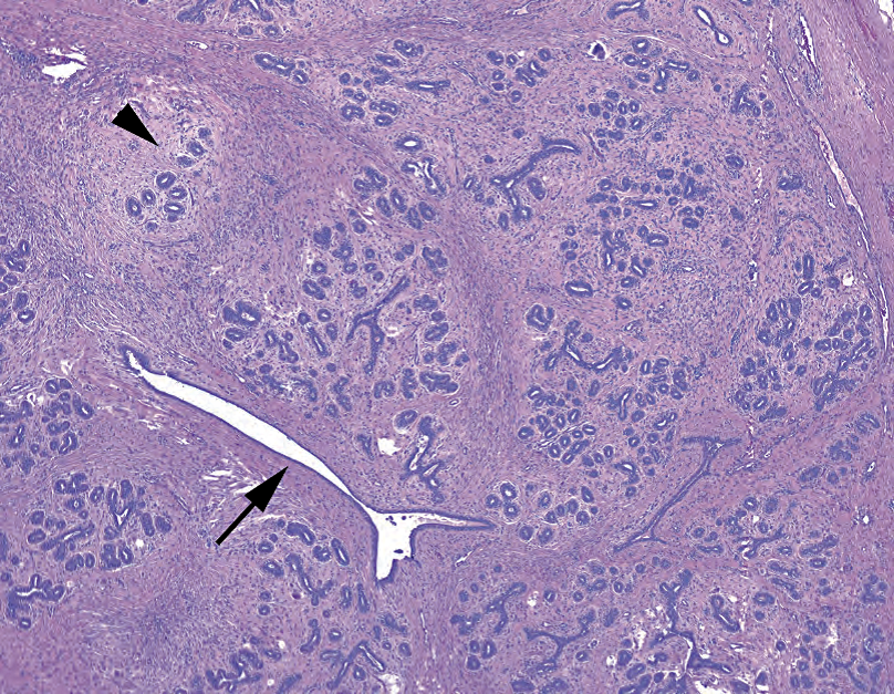
Figure 19.6. Fibroadenoma. At low power, the fibroadenoma is a well-circumscribed nodule. Within the lesion, the secretory lobules stand out in slightly edematous (pale) stroma (arrowhead), and the ducts are compressed into slit-like spaces (arrow) by the proliferative stroma.
图19.6 纤维腺瘤。低倍镜下,纤维腺瘤是一个边界清晰的结节。在病变内,分泌小叶位于淡染的轻度水肿的间质(箭头)中,间质增殖把导管挤压成狭缝状腔隙(箭)。
The phyllodes tumor is another biphasic lesion, which has a similar appearance but a much more cellular stroma that a fibroadenoma. The phyllodes (leaf-like) tumor is graded based on how aggressive the stromal growth pattern is and ranges from benign to malignant. This leaf-like pattern is often indicative of biphasic tumors with a very proliferative stroma and is seen in biphasic tumors of other organs.
叶状(像树叶)肿瘤是另一种双相病变,其外观与纤维腺瘤相似,但有更多细胞丰富的间质。根据间质生长模式的浸润性程度,把叶状肿瘤分级,范围从良性到恶性。这种叶状模式通常提示双相肿瘤伴非常增殖的间质,在其他器官的双相肿瘤中也可见。
Fat necrosis is evidence of a prior biopsy or other trauma. It can be hard, painful, calcified, or discolored. By clinical examination it may be very suspicious for malignancy. It is also very distracting in interpreting reexcision biopsies, where the prior surgery has left extensive fat necrosis. The key features (Figure 19.7) are as follows:
脂肪坏死是先前活检或其他创伤的证据。它可能是坚硬的、疼痛的、钙化的或变色的。临床检查可能高度怀疑恶性肿瘤。解读再次切除活检时,这可能非常分散注意力,因为先前的手术留下了广泛的脂肪坏死。主要特征(图19.7)如下:
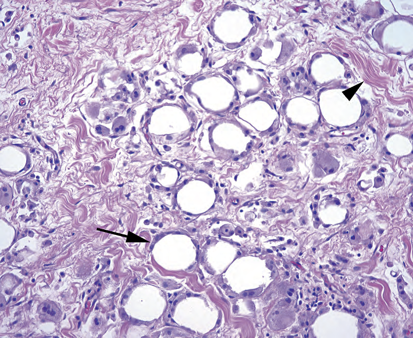
Figure 19.7. Fat necrosis. In an area of fat necrosis, secondary to trauma or surgery, the fat cells die but the globs of lipid remain. Foamy macrophages ring each dead fat cell (arrow), digesting the lipid; the spaces between the fat cells are filled in by fibrosis (arrowhead).
图19.7 脂肪坏死。在继发于创伤或手术的脂肪坏死区域,脂肪细胞死亡,但脂质空泡仍然存在。泡沫状巨噬细胞包围每个死亡的脂肪细胞(箭),消化脂质;脂肪细胞之间的空隙被纤维化填充(箭头)。
Disrupted and irregular fat cells
脂肪细胞紊乱,不规则
Foamy macrophages and giant cells
泡沫状巨噬细胞和巨细胞
Edema and hemosiderin
水肿和含铁血黄素
Acute inflammation
急性炎症
Fibrosis and calcification (in older lesions)
纤维化和钙化(陈旧病变)
导管内乳头状瘤(Intraductal Papilloma)
The papilloma is composed of proliferative but benign secretory and myoepithelial cells lining a branching arbor of fibrovascular cores (Figure 19.8). The lesion is usually found in the large distal ducts and can become fibrotic (sclerosing papilloma) or calcified with age. Rarely, carcinoma can arise in a papilloma.
乳头状瘤具有分支状纤维血管轴心,其上方被覆着增殖性但良性的分泌细胞(译注,导管细胞)和肌上皮细胞(图19.8)。病变通常位于远端大导管中,随着年龄的增长,病变可变为纤维化(硬化性乳头状瘤)或钙化。乳头状瘤内很少发生癌。
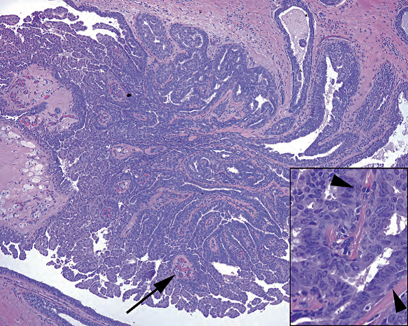
Figure 19.8. Intraductal papilloma. The branching structure fills a subareolar duct; smaller, more distal examples may be called micropapillomas. Although there is florid usual ductal hyperplasia, resulting in fusion of multiple branches of the papilloma, distinct fibrovascular cores are still visible (arrow). Inset: Along each fibrovascular core, you should still see myoepithelial cells (arrowheads), which differentiates this from a papillary carcinoma.
图19.8 导管内乳头状瘤。这种分支结构充满乳晕下大导管;较小的较远端导管内发生的病例可称为微小乳头状瘤。本例尽管有旺炽性导管增生,导致乳头状瘤的多个分支融合,但仍可见明显的纤维血管轴心(箭)。插图:沿着每个纤维血管轴心,仍然可见肌上皮细胞(箭头),这可以区分乳头状癌。
导管和小叶增殖性病变(Ductal and Lobular Proliferative Lesions)
This is the take-home point of the day: Deciding whether a lesion is ductal or lobular has nothing to do with whether you find it in a duct or a lobule. Lobular carcinoma in situ (LCIS) can fill a duct, and DCIS can invade a lobule, so there is no need to struggle to identify which structure you are looking at. Instead, ductal and lobular refer to distinct morphologic patterns of in situ or invasive carcinoma. They probably represent cancer pathways arising from a common cell type by two different mechanisms, analogous to the two cancer pathways in colon, but there are plenty of examples of “tweener” lesions (features of both) that are signed out as “mixed mammary carcinoma.”
今天的重点:理解“小叶”和“导管”这两个术语。确定某个病变是导管性还是小叶性,与你在导管内还是在小叶内发现它们无关。小叶原位癌(LCIS)可以填充导管,而导管原位癌(DCIS)可以侵入小叶,因此无需费劲去识别你所看到的结构。相反,导管和小叶是指原位癌或浸润性癌的不同形态模式。它们可能代表一种共同细胞类型通过两种不同机制的癌症途径,类似于结肠癌中的两种癌症途径,但也有大量“墙头草”病变(兼有导管和小叶特征)的病例,称为“混合性乳腺癌”。
普通型导管增生(usual ductal hyperplasia)
Benign hyperplasia of ductal-type epithelium (usual ductal hyperplasia) is common, whereas benign hyperplasia of lobular-type epithelium is not. However, both cell types can occur in an atypical proliferative phase (ADH and ALH), carcinoma in situ (DCIS and LCIS), and invasive carcinoma (IDC and ILC).
导管型上皮的良性增生(普通型导管增生)是常见的,而小叶型上皮的良性增生少见。然而,这两种细胞类型均可发生非典型增生(ADH和ALH)、原位癌(DCIS和LCIS)和浸润性癌(IDC和ILC)。
(译注:英文缩写ADH非典型导管增生,ALK非典型小叶增生,IDC浸润性导管癌,ILC浸润性小叶癌)
Usual ductal hyperplasia refers to a proliferation of cells within the ducts. The usual monolayer of cuboidal cells heaps up into mounds or even fills the ducts. Features of usual ductal hyperplasia include the following:
普通型导管增生是指导管内的细胞增殖。原先正常的单层立方形细胞堆积成小丘,甚至填满导管。普通型导管增生的特征包括:
The cells have an overall pale look; they are normochromic.
细胞整体上淡染;它们是正常染色的。
Cells appear jumbled, overlapping, or streaming and almost syncytial (Figure 19.9).
细胞显得杂乱、重叠或流水状排列,几乎是合胞体(图19.9)。
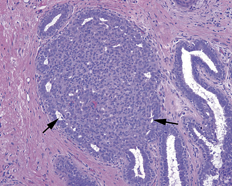
Figure 19.9. Florid usual ductal hyperplasia. The cellular proliferation entirely fills this duct, but the cell population is swirly and heterogeneous, with randomly overlapping nuclei. The peripheral ring of slit-like spaces (arrows), as though this clot of cells floated into the duct and stuck there, is very typical of usual ductal hyperplasia.
图19.9 旺炽性普通型导管增生。细胞丰富的增殖完全填满了这个导管,但细胞群呈漩涡状和异质性,细胞核随机重叠。周围有环状分布的狭缝状空隙(箭),好像这团细胞漂浮在导管中并粘在那里,这是非常典型的普通型导管增生的表现。
Heterogeneity (not to be confused with pleomorphism) is present. The nuclei, all bland with even chromatin and smooth nuclear membranes, range slightly in size and shape as though they were drawn by a sloppy artist.
存在异质性(不要与多形性混淆)。核都是形态温和,染色质均匀,核膜光滑,大小和形状稍有变化,就像马虎的画作。
Ducts may be filled with cells and may even have a cribriform look at low power, but on higher power the nuclei should be streaming, flowing parallel to the lumens, as opposed to polarizing perpendicularly (radially) around the lumen. Luminal spaces should be slit-like or irregular, not round, and may be “fuzzy” (due to apocrine secretions).
导管可能充满细胞,在低倍镜下甚至可能貌似筛状结构,但在高倍镜下,细胞核应为流水状,平行于管腔,而不是垂直于管腔周围(放射状)的极性。管腔应该是狭缝状或不规则的,而不是圆形的,并且管腔边缘可能是“模糊的”(由于顶浆分泌物)。
导管原位癌(ductal carcinoma in situ)
Ductal carcinoma in situ may be low grade, which is a homogeneous population of cells, or high grade, which is a pleomorphic population of cells. In low-grade DCIS, you should get the impression that there is a monotonous, clonal population of cells, with evenly spaced dark nuclei and distinct cell borders. High-grade DCIS, although it loses its monotonous look, should still have discrete nonoverlapping cells; it also may get very pink. Irregular nuclear borders, enlarged nuclei, and nucleoli are common. Patterns of DCIS include the following:
导管原位癌可以是低级别,含有均匀一致的细胞群,也可以是高级别,含有多形性细胞群。在低级别DCIS中,你的印象应该是:细胞群是单调的、单克隆的,具有均匀分布的深染核和清晰的细胞边界。高级DCIS虽然失去了单调的外观,但仍应具有离散的非重叠细胞;它也可能变得非常粉红色。常见核边界不规则、核增大和核仁是。DCIS的模式如下:
Cribriform: sharply punched-out round holes in the mass of cells, with cells lined up around the lumens like rosettes (Figure 19.10)
筛状:在细胞团中尖锐地冲凿出圆孔,细胞排列在管腔周围,像菊形团(图19.10)
Solid: a solid sheet of monotonous cells
实性:实性成片的单调细胞
Comedo: a rim of malignant, usually high grade, cells with central necrosis (see Figure 19.10)
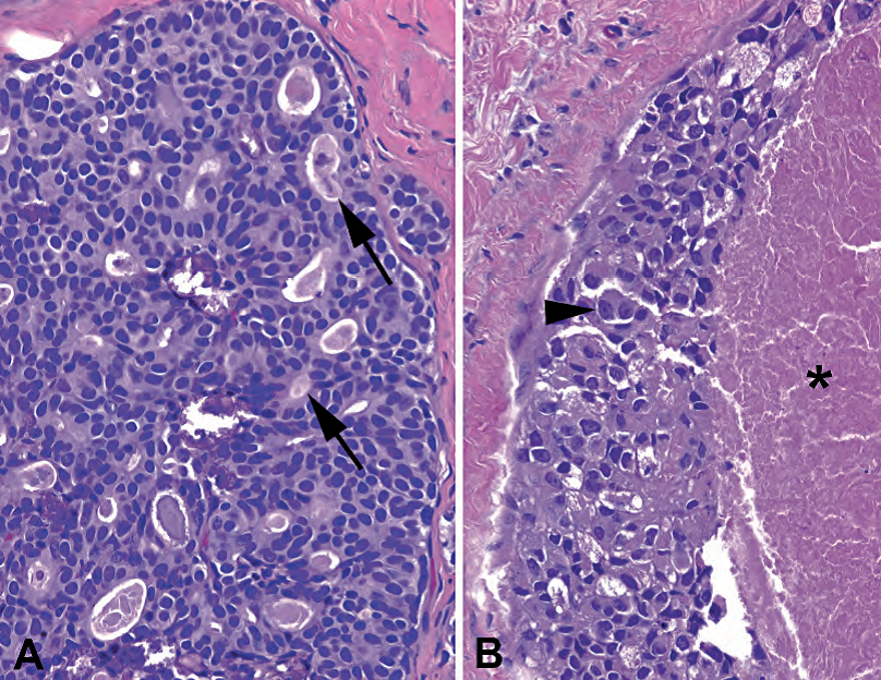
Figure 19.10. Ducal carcinoma in situ (DCIS). (A) In low-grade DCIS, the cells are monotonous, uniform, and largely nonoverlapping, and they form cribriform duct spaces with the cells polarized around the tiny lumens (arrows). (B) In high-grade DCIS, the cells have lost their monotony and are instead pleomorphic, some with prominent nucleoli (arrowhead). At the center of the dilated duct there is necrosis (asterisk), indicating comedo-type DCIS.
图19.10 导管原位癌(DCIS)。(A)在低级别DCIS中,细胞单调、均匀,基本上不重叠,它们形成筛状导管腔,围绕微小管腔的细胞有极性(箭)。(B)在高级别DCIS中,细胞已失去了单调性,而是多形性,一些细胞具有显著核仁(箭头)。扩张导管的中央有坏死(星号),提示粉刺型DCIS。
粉刺:中央坏死,边缘有一圈恶性细胞,通常为高级别(见图19.10)
Micropapillary: top-heavy lollipop protrusions into the lumen, without true fibrovascular cores; must also have cellular monotony as above.
微乳头状:顶部增大的棒棒糖,突向管腔内,没有真正的纤维血管轴心;还必须具有上述的细胞单调性。
Remember that DCIS by definition has not crossed the basement membrane, and the outer myoepithelial layer remains intact. Ductal carcinoma in situ is treated as a precursor to malignancy, and the treatment goal is total excision. Therefore, on anything but a core biopsy, you must document its distance from each margin (adequate clearance is in the eye of the beholder, but most accept 2 mm).
记住,根据定义,DCIS没有穿过基底膜,并且外面的肌上皮层保持完整。导管原位癌视为恶性肿瘤的前驱病变,治疗目标是完整切除。因此,除了粗针穿刺活检之外的任何标本,必须记录DCIS至每个边缘的距离(在观察者眼中有足够的距离,但大多数接受2mm为安全切缘)。
非典型导管增生(atypical ductal hyperplasia)
Do not expect to get comfortable with the diagnosis of atypical ductal hyperplasia until you have mastered usual ductal hyperplasia and DCIS. Atypical ductal hyperplasia falls somewhere in between and has no definitive criteria other than “has some but not all of the features of DCIS.” This diagnosis is also used in the setting of a single focus of apparent low-grade DCIS (nuclear grade 1 of 3) measuring less than 3 mm. In a core biopsy, atypical ductal hyperplasia is code for “get me more tissue.” Features that can push you to DCIS in a tiny focus include high-grade nuclei and/or necrosis.
尚未掌握普通型导管增生和DCIS的初学者,不要满足于诊断非典型导管增生。非典型导管增生介于两者之间,除了“具有DCIS的部分特征而不是全部特征”之外,没有明确的标准。该诊断也适用于单个病灶的明显低级别DCIS(3级核分类中的1级),但测量值小于3mm。在粗针穿刺活检中,诊断非典型导管增生将促使临床进行扩大切除术。在微小病灶中,可能把你推向DCIS诊断的特征包括高级别核和/或坏死。
浸润性导管癌(invasive ductal carcinoma)
Invasive ductal carcinoma is invasive carcinoma arising from a DCIS lesion, and therefore the cells of invasive ductal look similar to those you see in DCIS. In its most common form, invasive ductal carcinoma is the cancer formerly known as scirrhous, so called because of the dense desmoplastic reaction generated. It is eye-catching even to the untrained eye, as a large cellular lesion with ugly cells, radiating outward in a stellate and decidedly un-TDLUlike shape (Figure 19.11). The cells are large, with large pleomorphic nuclei and substantial pink cytoplasm. Necrosis and mitoses are common. Nests of tumor cells can imitate ducts or tubules in the stroma, or acquire large necrotic centers like comedo-DCIS. For this reason it is sometimes hard to tell invasive carcinoma from DCIS or even benign tubules. Stains for myoepithelial borders are helpful here: invasive cancer does not have any.
浸润性导管癌是来自DCIS病变的浸润性癌,因此浸润性导管癌的细胞看起来类似于DCIS细胞。最常见的一种浸润性导管癌以前称为硬癌,因为它产生致密的促结缔组织增生反应。即使没有接受过病理训练,它也是引人注目的,因为它是巨大的细胞丰富的病变,细胞丑陋,向外辐射,呈星芒状,明显不像TDLU结构(图19.11)。细胞大,有大的多形核和大量粉红色细胞质。常见坏死和核分裂。肿瘤细胞巢可能貌似间质中的导管或小管,或者像粉刺型DCIS的大片坏死中心。因此,有时很难区分浸润性癌和DCIS甚至良性小管。肌上皮标记物免疫染色有助于鉴别:浸润性癌的边缘没有任何残留的肌上皮细胞。
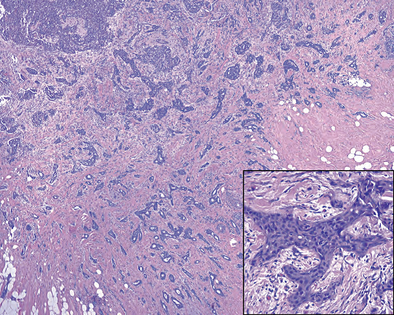
Figure 19.11. Infiltrating ductal carcinoma. At low power, the irregular border of the lesion is evident, with small angular tubules radiating outward into the fat. Grossly, this lesion would have a stellate appearance, and the dense stromal reaction would make the lesion very hard. Inset: The irregularly shaped nests of tumor cells create a desmoplastic stromal reaction, which is a combination of edema (white space) and fibrosis (pink collagen).
图19.11 浸润性导管癌。低倍镜下,病变边界明显不规则,小的成角小管向外辐射,进入脂肪中。大体上,该病变应当呈星芒状,致密的间质反应使病变非常坚硬。插图:不规则形状的肿瘤细胞巢产生促结缔组织增生性间质反应,表现为水肿(白色空隙)和纤维化(粉红色胶原)的组合。
Variants of ductal carcinoma include the following (the first five variants have a generally better prognosis than ductal carcinoma NOS):
以下列举几种特殊型浸润性导管癌(前五种通常比浸润性导管癌NOS预后更好):
(译注:NOS(not otherwise specified),非特指。NST(no specialtype),非特殊型。二者是同义词,更简单说“普通型”)
Tubular: a very well-differentiated cancer composed entirely of cytologically bland small angular tubules (Figure 19.12)
小管癌:一种分化非常好的癌,完全由细胞学温和的成角状小管组成(图19.12)
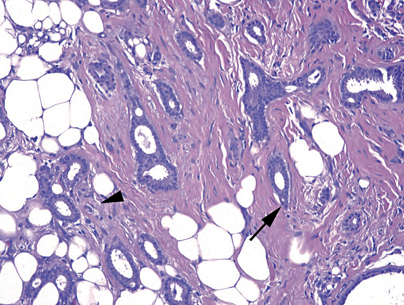
Figure 19.12. Tubular carcinoma. Well-formed tubules with pointed ends (arrow) and round, monotonous cells infiltrate through the stroma and fat. The myoepithelial layer is absent, both on H&E stain and by immunostain, and there is a subtle desmoplastic reaction around some of the tubules (arrowhead).
图19.12 小管癌。形状完好的小管,一端尖细(箭),圆形、单调的细胞浸润间质和脂肪。在HE染色和免疫染色中,肌上皮层均不存在,一些小管周围有轻微的促结缔组织增生反应(箭头)。
Cribriform: similar to tubular, but with cribriform structures instead of tubules
筛状癌:类似小管癌,但有筛状结构而不是小管
Mucinous or colloid: characterized by pools of mucin and floating fragments of neoplastic epithelium (Figure 19.13)
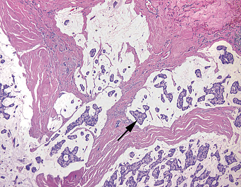
Figure 19.13. Mucinous carcinoma. Pools of extruded mucin dissect into the stroma. Although this can occur in benign mucocele-like lesions, the presence of floating clumps of cells (arrow) is diagnostic of mucinous, or colloid, carcinoma.
图19.13 粘液癌。粘液渗入间质形成粘液池。虽然良性粘液囊肿样病变也可能发生粘液池,但漂浮的细胞团(箭)是粘液癌或胶样癌的诊断性特征。
黏液癌或胶样癌:特征是黏液池及其中漂浮的肿瘤上皮片段(图19.13)
Medullary: a well-circumscribed but paradoxically ugly group of cells, with a dense lymphocytic infiltrate
髓样癌:肿瘤边界清晰,但矛盾的是,细胞学丑陋,并有致密的淋巴细胞浸润
Adenoid cystic carcinoma: a biphasic tumor of epithelial and myoepithelial cells, identical to the salivary gland tumor of the same name
腺样囊性癌:上皮细胞和肌上皮细胞组成的双相肿瘤,形态学等同于涎腺同名涎腺的腺样囊性癌
Metaplastic: a tumor in which there is a mesenchymal or spindle-cell component, such as cartilage, bone, or frank sarcoma, with prognosis depending on grade
化生性癌:肿瘤中含有间叶性成分或梭形细胞成分,如软骨、骨或明显肉瘤,其预后取决于分级
小叶原位癌(lobular carcinoma in situ)
In lobular carcinoma in situ, lobular cells, when they begin to proliferate, take on a characteristic appearance. They are homogeneous, like DCIS cells, but they have a round fried-egg shape, with a pale cytoplasm, discrete borders, and a central round nucleus (Figure 19.14). Intracytoplasmic vacuoles, even signet-ring cells, are also common. In LCIS, these cells should fill and expand the lobules, appearing at low power like a very circumscribed stippled space (such as the texture of newspaper photos under a magnifying glass). Lobular carcinoma in situ retains its bland cytology right through to invasive carcinoma.
在小叶原位癌中,增殖的小叶细胞呈现特征性细胞形态学。像低级别DCIS细胞一样,它们是同质的,但它们呈圆形的煎鸡蛋形状,细胞质淡染,细胞边界不相连,核圆,居中(图19.14)。胞质内空泡,甚至印戒细胞也很常见。在LCIS中,这些细胞应该填充并扩张小叶,低倍镜下,就像一个边界非常清楚的腔隙(如放大镜下报纸照片的纹理)。小叶原位癌保留其形态温和的细胞学直到浸润性癌。
(译注:这段文字描述不太好。小叶原位癌的细胞学特征:非粘附性低级别均质性小细胞;可有胞质空泡,使核偏位,直至印戒细胞样)
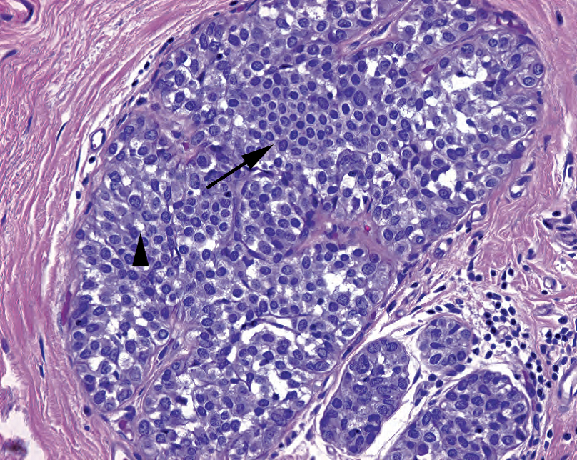
Figure 19.14. Lobular carcinoma in situ. The lobule is distended by a population of monotonous cells with distinct cellular borders and small round nuclei (arrow). As the lesion expands, the noncohesive cells will begin to fall apart. Cytoplasmic vacuoles (arrowhead) are typical of lobular carcinoma cells both in situ and invasive.
图19.14 小叶原位癌。小叶充满一群单形性细胞,具有明显细胞边界和小圆核(箭)。随着病变的扩大,非粘附性细胞开始分离。细胞质空泡(箭头)是原位和浸润性小叶癌细胞的典型特征。
Lobular carcinoma in situ is often multifocal and bilateral, and its progression to cancer is not considered inevitable or predictable. As a result, excision is not the goal of treatment, and so its presence at a margin is not usually noted.
小叶原位癌通常是多灶性和双侧性,它并非不可避免地可预测地进展为癌。因此,治疗目标不是切除,因此它在切缘出现时通常不需要注明。
Lobular carcinoma in situ is an incidental finding. It does not form masses or calcify (usually). Atypical lobular hyperplasia is generally code for “I’m really worried about LCIS but cannot quite get there.” Like atypical ductal hyperplasia, atypical lobular hyperplasia does not have consistently agreed-upon criteria.
小叶原位癌是一种偶然发现。通常不会形成肿块或钙化。非典型小叶增生通常意味着“我真的很担心LCIS,但不能直接诊断”。与非典型导管增生一样,非典型小叶增生没有一致的标准。
E-cadherin is a cell surface molecule that helps cells stick together. Lobular lesions lose expression of E-cadherin and therefore begin to appear very discohesive. You can imagine that this nonsticky surface enables the invasive lobular cells to slip through the stroma as single cells, and that is exactly what they do. Stains for E-cadherin can help to sort out LCIS (negative) from DCIS (positive) in a core biopsy specimen, as low-grade DCIS can resemble LCIS.
E-钙粘蛋白是一种帮助细胞粘附在一起的细胞表面分子。小叶病变失去E-钙粘蛋白的表达,因此显得粘附性很差。你可以想象,这种不粘附的表面使浸润性小叶细胞以单个细胞的形式滑过间质,事实正是如此。E-钙粘蛋白染色有助于区分粗针穿刺活检标本中的LCIS(阴性)和DCIS(阳性),因为低级别DCIS可能类似于LCIS。
浸润性小叶癌(invasive lobular carcinoma)
The cells of invasive lobular carcinoma look similar to those of LCIS. They are small uniform cells with bland round nuclei, pale cytoplasm, and a sometimes plasmacytoid shape with an eccentric mucin vacuole. Because of their normochromic nuclei and lack of malignant cytology, they are identified by the way they slip through the stroma. They line up as single file lines or as concentric rings around ducts and do not cause an appreciable desmoplastic response (Figure 19.15). They are sneaky and scary, and you have not ruled out lobular until you have looked closely at 10× or 20×. A cytokeratin stain can highlight the individual cells, as everything else in the stroma should be negative.
浸润性小叶癌的细胞类似于LCIS的细胞。细胞小而均匀,核温和圆形,胞质淡染,有时呈浆细胞样,带有偏心的粘液空泡。由于核染色正常,并且缺乏恶性细胞学特征,因此通过它们滑过间质的方式来识别。它们单行排列,或同心环状排列在导管周围,不会引起明显的促结缔组织增生反应(图19.15)。它们鬼鬼祟祟、令人恐惧,在仔细观察10×或20×之前,你不能排除小叶癌。CK染色可以突出显示单个细胞,因为间质中的其他任何东西都应该是阴性的。
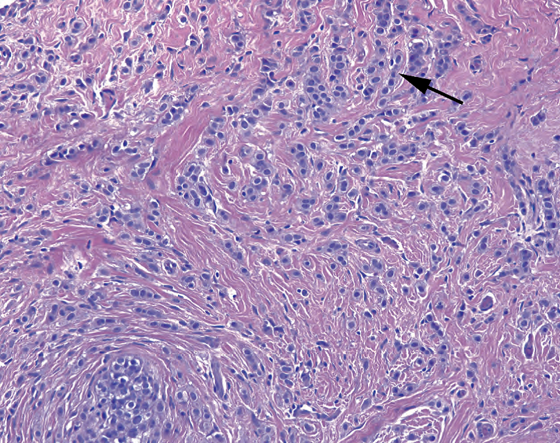
Figure 19.15. Invasive lobular carcinoma. The same cells as in Figure 19.14 are seen here invading through the stroma. They often form single file lines (arrow) but may also be seen as single cells or concentric circles around a duct. In some cases there is little to no desmoplastic stromal reaction, making the lesion difficult to palpate or detect.
图19.15 浸润性小叶癌。与图19.14形态学相同的细胞浸润间质。它们通常呈单行线状排列(箭),但也可见单个细胞,或围绕导管的同心圆。在某些病例中,几乎没有或完全没有促结缔组织增生性间质反应,使病变难以触诊或检出。
Elston分级(Elston grade)
Invasive epithelial carcinomas must be given an Elston grade (A.K.A Nottingham grade, or Elston-Ellis modification of Scarff-Bloom-Richardson) when diagnosed in a lumpectomy or mastectomy. The Elston grade is the pathologic assessment of the tumor’s aggressiveness; the stage is diagnosed separately by features such as size and local extent. The Elston grade takes into account three prognostic factors:
肿块切除术或乳房切除术诊断的浸润性癌,必须给予Elston分级(又称诺丁汉分级,或Scarff Bloom-Richardson修订的Elston-Ellis分级)。Elston分级是肿瘤侵袭性的病理评估;而分期是根据肿瘤大小和局部浸润范围等特征而单独诊断的。Elston分级考虑了三个预后因素:
Tubule formation (the more tubule formation, the lower the score)
小管形成(小管形成越多,得分越低)
Mitotic rate (the more mitoses, the higher the score)
核分裂率(核分裂越多,得分越高)
Pleomorphism (the more pleomorphic the nuclei, the higher the score)
多形性(细胞核多形性越强,分数越高)
Each characteristic is scored from 1 to 3, and then all are added, to give you a range of 3 to 9. For details on scoring, see your favorite surgical pathology text; in the beginning, just learn to look for these three features. Pleomorphism, especially, is a fairly subjective criterion that takes some experience to judge.
每个特征的得分从1到3,然后全部相加,得到3到9的范围。有关评分的详细信息,请参阅你最喜欢的外科病理学教材;在开始时,只需学习寻找这三个特性。尤其是多形性,是一个相当主观的标准,需要一些经验来判断。
乳头状病变的命名(Papillary Nomenclature)
Papillary lesions in the breast represent a confusing area. Here is the nutshell.
乳腺乳头状病变是容易混淆的地方。简介如下。
A papilloma is a benign lesion with papillary architecture. The fibrovascular cores, and the surrounding duct, are lined by myoepithelial cells. Within a papilloma, you can get usual or atypical ductal hyperplasia or DCIS, all of which are diagnosed as “arising in a papilloma.” You should still have myoepithelial cells around the perimeter.
乳头状瘤是一种具有乳头状结构的良性病变。纤维血管轴心和周围的导管都有肌上皮细胞排列。在乳头状瘤中,可有UDH、ADH或DCIS,所有这些都被诊断为“发生在乳头状瘤中”。周围仍然应该有肌上皮细胞。
Within the DCIS family, there are several architectural types: micropapillary (epithelial projections without fibrovascular cores), papillary (epithelial projections with fibrovascular cores), and solid papillary (a solid ball of cells with residual entombed fibrovascular cores). None of these necessarily has anything to do with a papilloma. All usually have intact myoepithelial cells around the outside. All may be multifocal processes in the breast.
在DCIS家族中,有几种结构类型:微乳头状(无纤维血管轴心的上皮突起)、乳头状(有纤维血管轴心的上皮突起)和实性乳头状(有残余埋陷的纤维血管轴心的实性细胞团)。这些结构都不一定与乳头状瘤有关。所有这些结构的外周通常都有完整的肌上皮细胞。所有这些结构都可能是乳房中的多灶性病变。
Papillary carcinoma is a specific type of carcinoma with a papillary architecture, homogeneous columnar cells, and a circumscribed profile, as though it once grew in a duct. It should be a single discrete lesion. The fibrovascular cores have no myoepithelial cells. The myoepithelial stains may also be negative around the perimeter, but it is still not really considered a true invasive carcinoma. It may be called intracystic or encysted papillary carcinoma to get this point across.
乳头状癌是一种特殊类型的癌,具有乳头状结构、均匀的柱状细胞和边界清楚的轮廓,就像它曾经生长在导管中一样。它应该是单个的孤立性病变。纤维血管轴心没有肌上皮细胞。肌上皮染色也可能在周围呈阴性,但它仍然不是真正的浸润性癌。这种可以称为囊内乳头状癌或包裹性乳头状癌。
化生癌的多面性(The Many Faces of Metaplastic Carcinoma)
Numerous morphologies get lumped under the term metaplastic carcinoma and hence the struggle to learn to recognize it. You may see this diagnosis applied to the following entities:
许多形态都被归为化生癌,因此很难学会识别它。你可以看到此诊断适用于以下实体:
Squamous carcinoma: a ductal carcinoma with prominent squamous differentiation (and technically a form of metaplastic carcinoma)
鳞状细胞癌:一种具有显著鳞状分化的导管癌(技术上是化生癌的一种形式)
Low-grade spindle cell carcinoma: can masquerade as a hypercellular stroma, but the spindle cells should stain for cytokeratins (especially high-molecular-weight cytokeratins such as 34bE12 [CK903])
低级别梭形细胞癌:可以伪装成细胞丰富的间质,但梭形细胞应表达CK(特别是高分子量细胞角蛋白,如34bE12[CK903])(译注:实际上CK5/6和p63更有用更常用)
High-grade carcinoma with spindle cell features: should also be cytokeratin positive
具有梭形细胞特征的高级别癌:也应为CK阳性
Any carcinoma with coexisting sarcoma, such as chondrosarcoma or osteosarcoma (The carcinoma component will be cytokeratin positive, the sarcoma usually will not. In another organ this would be called a carcinosarcoma.)
任何与肉瘤共存的癌,如软骨肉瘤或骨肉瘤(癌成分为CK阳性,肉瘤通常阴性。在其他器官中称为癌肉瘤。)
The differential diagnosis for entities that are spindly and malignant also includes malignant phyllodes tumor and primary or metastatic sarcoma.
梭形细胞恶性肿瘤的鉴别诊断还包括恶性叶状肿瘤和原发性或转移性肉瘤。
来源:
The Practice of Surgical Pathology:A Beginner’s Guide to the Diagnostic Process
外科病理学实践:诊断过程的初学者指南
Diana Weedman Molavi, MD, PhD
Sinai Hospital, Baltimore, Maryland
ISBN: 978-0-387-74485-8 e-ISBN: 978-0-387-74486-5
Library of Congress Control Number: 2007932936
© 2008 Springer Science+Business Media, LLC
仅供学习交流,不得用于其他任何途径。如有侵权,请联系删除。
本站欢迎原创文章投稿,来稿一经采用稿酬从优,投稿邮箱tougao@ipathology.com.cn
相关阅读
 数据加载中
数据加载中
我要评论

热点导读
-

淋巴瘤诊断中CD30检测那些事(五)
强子 华夏病理2022-06-02 -

【以例学病】肺结节状淋巴组织增生
华夏病理 华夏病理2022-05-31 -

这不是演习-一例穿刺活检的艰难诊断路
强子 华夏病理2022-05-26 -

黏液性血性胸水一例技术处理及诊断经验分享
华夏病理 华夏病理2022-05-25 -

中老年女性,怎么突发喘气困难?低度恶性纤维/肌纤维母细胞性肉瘤一例
华夏病理 华夏病理2022-05-07


共0条评论