外科病理学实践:诊断过程的初学者指南 | 第6章 食管
第6章 食管(Esophagus)
The esophagus is composed of a nonkeratinized squamous epithelium overlying a lamina propria and thin muscularis mucosa. The submucosa contains lymphatics and mucous glands with cuboidal-lined ducts running up to the luminal surface. Under the submucosa is muscularis propria (skeletal muscle proximally, smooth muscle distally), surrounded by the adventitia, which is continuous with mediastinum.
食管由非角化鳞状上皮组成,下方是固有层和薄的粘膜肌层。粘膜下层含有淋巴管和粘液腺,其导管呈立方形排列,向上延伸到管腔表面。粘膜下层之下为固有肌层(近端为骨骼肌,远端为平滑肌),由外膜包围,外膜与纵隔相连。
Most esophageal biopsies are performed on patients with symptoms of reflux or dysphagia and often the goal is to rule out Barrett’s esophagus, a glandular metaplasia that puts the patient at increased risk for adenocarcinoma. Other common findings include reflux changes in the squamous epithelium, ulcers, or infection. Squamous dysplasia is actually uncommon.
大多数食管活检是针对有反流或吞咽困难症状的患者,其目的通常是排除Barrett食管,这是一种腺体化生,会增加患者发生腺癌的风险。其他常见的发现包括鳞状上皮的反流改变、溃疡或感染。鳞状细胞异型增生实际上并不常见。
On low power, survey the epithelium. A normal biopsy specimen (Figure 6.1) will have a bland pink squamous epithelium and scant submucosa; muscularis propria should not be present. Occasional lymphocytes in the epithelium are typical (so-called squiggle cells because of their stretched-out appearance). The epithelium should not be interrupted or undermined by gastric-type glands, although salivary-like mucous glands are okay.
在低倍镜下,仔细观察上皮。正常活检标本(图6.1)会有形态温和的淡粉色鳞状上皮和少量粘膜下层;固有肌层不应存在。上皮中偶尔出现淋巴细胞,是典型的或正常的(由于其伸展的外观而称为蠕动细胞)。上皮不应该被胃型腺体打断或破坏,尽管涎腺样粘液腺是可以的。
Within the squamous epithelium, look for the following:
在鳞状上皮内,观察以下情况:
Basal cell hyperplasia (an increase over the normal three-cell layer) (Basal cells are the deepest layer of squamous cells and are the regenerative cell layer. They are defined by their closely packed nuclei: if you cannot squeeze a new nucleus between two existing nuclei, they are basal cells.)
基底细胞增生(超过正常的三层细胞)(基底细胞是鳞状细胞的最深层,是生发细胞层。它们具有紧密排列的细胞核:如果你不能在两个既有核之间挤进去一个新核,它们就是基底细胞。)
Elongated vascular papillae (over two thirds of the thickness of the epithelium)
细长的血管乳头(超过上皮厚度的三分之二)
Balloon cell change of epithelium (an accumulation of glycogen in the cytoplasm)
上皮的气球细胞改变(细胞质中糖原的积累)
Intraepithelial neutrophils or eosinophils
上皮内中性粒细胞或嗜酸性粒细胞
Erosions, fibrinopurulent exudate, granulation tissue
糜烂、纤维素渗出物、肉芽组织
Columnar cell mucosa or glands
柱状细胞粘膜或腺体
反流改变(Reflux Changes)
A very common finding on biopsy is reflux esophagitis, which consists of the first four features in the preceding list (Figure 6.2). Not all features need be present in every case and typically are not. Severe cases may progress to erosions and ulcerations. The inflammation in reflux is mainly lymphocytic, but eosinophils may be seen; a very high number of eosinophils (or clustering of three or more) may indicate eosinophilic esophagitis. Stylistically, we often use the phrase “reactive epithelial changes” to describe changes that have some, but not all, of the features of full-blown reflux esophagitis.
反流性食管炎是很常见的活检发现,它包括前面列表中的前四个特征(图6.2)。并非每个病例都会出现所有特征,通常情况下并非如此。严重的病例可能发展为糜烂和溃疡。反流性炎症主要为淋巴细胞性,但可能见到嗜酸性粒细胞;大量嗜酸性粒细胞(或≥3个成簇聚集)可能提示嗜酸性食管炎。在措辞上,我们经常使用“反应性上皮改变”来描述只有完全反流性食管炎的部分特征(但不是全部)的改变。
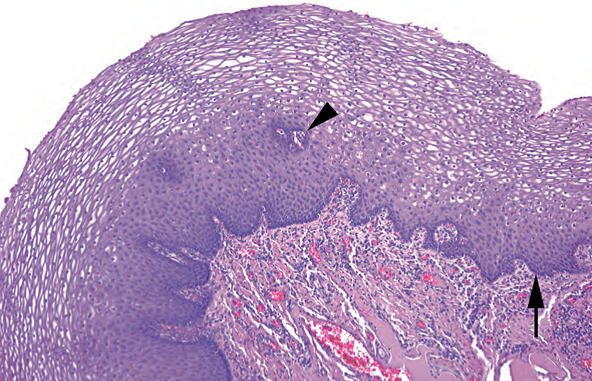
Figure 6.1. Normal esophageal mucosa. The basal layer (arrow) is seen as a crowded and blue layer at the base. The cells mature into flat nonkeratinizing squamous cells with small nuclei; the clear cytoplasm seen here is glycogen. Vascular pegs penetrate into the epithelium (arrowhead). The vascular lamina propria is visible below the basal layer.
图6.1.正常食管粘膜。基底层(箭号)看起来是底部一层密集的蓝色。细胞成熟为扁平、非角质化鳞状细胞,核小;细胞质透明,含有糖原。血管钉突伸入上皮(箭头)。在基底层下方可见血管丰富的固有层。
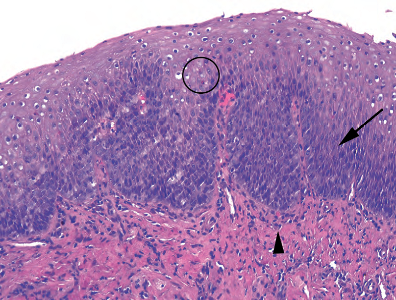
Figure 6.2. Reflux esophagitis. Compare this inflamed epithelium to the normal mucosa in 图6.2.反流性食管炎。表现为炎症性上皮,与图6.1正常粘膜相比较。基底层的厚度增加(箭号),使上皮看起来更致密更蓝。炎症细胞散在分布,包括嗜酸性粒细胞(圆圈)和淋巴细胞(箭头)。
中性粒细胞(Neutrophils)
A prominent neutrophilic infiltrate points more to an infection or acute injury rather than reflux. Look at the periodic-acid Schiff (PAS) stain to find Candida organisms (pseudohyphae and yeast forms in the epithelium or exudate; Figure 6.3). They may be very numerous or extremely scanty. Luminal squamous debris is another hint to look closely for Candida. Candidal infection is typically associated with a superficial neutrophilic infiltrate and parakeratosis (surface squames that are too red and have retained nuclei); however, some cases have almost no inflammation and little epithelial changes.
明显的中性粒细胞浸润更提示感染或急性损伤,而不是反流。观察PAS染色,查找念珠菌(上皮或渗出液中的假菌丝和酵母形式;图6.3)。它们可能非常多,也可能非常稀少。管腔鳞状碎屑也提示念珠菌感染,需仔细查找。念珠菌感染通常伴随表面中性粒细胞浸润和角化不全(表面鳞片太红且保留细胞核);然而,有些病例几乎没有炎症,上皮改变很小。
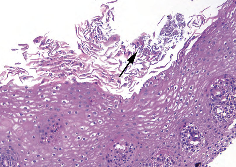
Figure 6.3. Candida. Tiny purple yeasts and pseudohyphae (arrow) are seen among the squamous debris at the surface of the epithelium. This is a hematoxylin and eosin stain; the yeasts are magenta on periodic-acid Schiff stain. Note that sometimes there is not a significant neutrophilic response.
图6.3.念珠菌。HE染色,上皮表面鳞状碎屑中可见微小的紫色酵母和假菌丝(箭头)。酵母在PAS染色上呈洋红色。注意,有时没有明显的中性粒细胞反应。
溃疡(Ulcers)
Ulcers can be caused by severe reflux or chemical injury (especially pill esophagitis; polarize to look for pill fragments), radiation (should be accompanied by necrosis and bizarre atypia), or infection, or they can be idiopathic (particularly in acquired immunodeficiency syndrome). Viral esophagitis is rare but more common in the immunosuppressed. Herpes simplex virus and cytomegalovirus cause inflamed, punched-out ulcers.
溃疡的病因包括严重反流或化学损伤(尤其是药片食管炎;用偏振光寻找药片碎屑)、辐射(应伴有坏死和奇异形非典型性)或感染,也可为特发性溃疡(尤其获得性免疫缺陷综合征)。病毒性食管炎罕见,但在免疫抑制患者较常见。单纯疱疹病毒和巨细胞病毒引起炎症、穿孔溃疡。
Herpes simplex virus infects epithelial cells. This is best seen on intact squamous mucosa adjacent to the ulcer, typically causing multinucleation with nuclear molding and glassy chromatin.
单纯疱疹病毒感染上皮细胞。这在邻近溃疡的完整鳞状粘膜上最为明显,通常导致多核伴核镶嵌和毛玻璃样染色质。
Cytomegalovirus infects mesenchymal cells (fibroblasts, endothelial, etc.) at the ulcer base. Cytomegalovirus infection usually manifests with intranuclear and cytoplasmic red/purple inclusions that render the cells gigantic (cyto-megalo-virus), and thus 10× is a good objective to scan with.
巨细胞病毒感染溃疡底部的间叶细胞(成纤维细胞、内皮细胞,等)。巨细胞病毒感染通常表现为核内和细胞质内的红色/紫色内含体,使细胞变得巨大(巨细胞病毒因此得名),因此10倍镜下扫描容易观察到。
The hematoxylin and eosin (HE) slide is the best place to find the inclusions, but immunostains may help if clinical suspicion is high and HE is not definitive (see Chapter 3 for images of viral inclusions).
HE切片是最容易发现包涵体,但如果临床怀疑但HE染色不确定,免疫染色可能会有帮助(见第3章中病毒包涵体的图像)。
柱状上皮(Columnar Epithelium)
粘膜活检材料中,偶尔可见类似于涎腺或十二指肠Brunner腺的粘膜下粘液腺的聚集。更常见的是看到这些腺体的导管。“食管”活检标本中的胃型上皮(小凹表面上皮和下方的特化胃腺,通常是泌酸腺)可能是无意中从近端胃或食管裂孔疝中取出的组织。出现贲门粘膜时要注意,但并不等同于Barrett的诊断(见后面的讨论)。上皮下粉紫色腺泡细胞的聚集,类似于正常胰腺,实际上可能就是正常胰腺(称为胰腺化生或异位)。
Collections of submucosal mucous glands resembling salivary glands or Brunner’s glands of duodenum are occasionally seen in mucosal biopsy material. It is more common to see the ducts from these glands. Gastric-type epithelium (foveolar surface epithelium and underlying specialized gastric glands, usually oxyntic) in an “esophagus” biopsy specimen may represent tissue inadvertently taken from proximal stomach or hiatal hernia. The presence of cardiac mucosa should be noted but does not equal a diagnosis of Barrett’s esophagus (see later discussion). Collections of pink-purple acinar cells beneath the epithelium, resembling normal pancreas, may in fact be normal pancreas (called pancreatic metaplasia or heterotopia).
杯状细胞(Goblet Cells)
In the tubular esophagus, the presence of goblet cells (bulbous epithelial cells that are indigo-blue on PAS/alcian blue (AB) stain, clear-to-pale-blue on HE; Figure 6.4) in glandular mucosa, otherwise known as intestinal metaplasia, indicates Barrett mucosa of the distinctive type (“Barrett’s esophagus”). There are two caveats to be considered. First, intestinal metaplasia can also occur in the true cardia of the stomach as a response to inflammation. Therefore, if the location where the biopsy tissue came from is not entirely clear, some pathologists will sign out apparent Barrett’s as “consistent with Barrett’s mucosa if the biopsy was taken from tubular esophagus.” Second, not all that stains blue with the PAS/AB is a goblet cell. Some gastric-type foveolar cells, especially at the squamocolumnar junction, will stain blue (so-called tall blues), so, to be a genuine goblet it has to stain blue and have goblet cell morphology (bulbous outline of goblet cells vs. the elongated foveolar cells).
在管状食管中,腺粘膜中出现杯状细胞(PAS/AB染色呈靛蓝色的球状上皮细胞,HE染色为透明至淡蓝色;图6.4),也称为肠化生,提示特殊类型的Barrett粘膜(Barrett食管)。需要注意两点。首先,作为对炎症的反应,肠化生也可能发生在真正的胃贲门。因此,如果活检组织来源的部位不完全清楚,一些病理医师会将明显的Barrett食管签发为“如果活检来自管状食管,则符合Barrett's粘膜”。其次,并非所有PAS/AB染色呈蓝色的都是杯状细胞。一些胃型小凹细胞,特别是在方柱连接处,会染上蓝色(所谓的高蓝色),因此,要成为真正的杯状细胞,它必须染上蓝色并具有杯状细胞形态(杯状细胞的球状轮廓与细长的小凹细胞)。
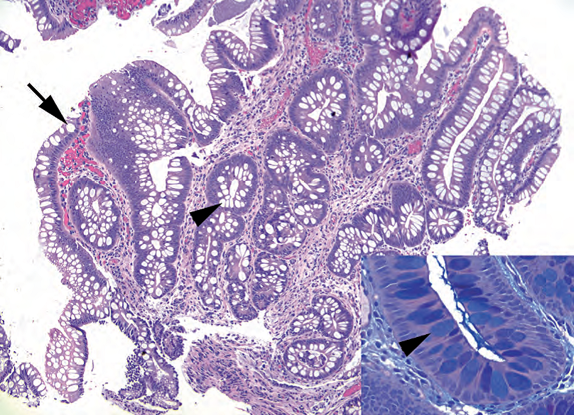
Figure 6.4. Goblet cells in Barrett’s esophagus. The presence of columnar epithelium with goblet cells indicates Barrett’s esophagus. Goblet cells are round cells that appear clear on hematoxylin and eosin stain and are typically flanked by the purplish absorptive-type cells. Back-to-back mucinous cells resembling a row of teeth are more likely to be gastric foveolar epithelium. Goblet cells may be present at the surface (arrow) or in deep glands (arrowhead). Inset: A periodic-acid Schiff/alcian blue stain confirms the goblet cells, which stain indigo blue (arrowhead).
图6.4.Barrett食管中的杯状细胞。出现含有杯状细胞的柱状上皮提示Barrett食管。杯状细胞呈圆形,HE染色呈透明胞质,两侧通常为紫色吸收型细胞。类似于一排牙齿的背靠背的粘液细胞更可能是胃小凹上皮。杯状细胞可能出现在表面(箭号)或深腺体(箭头)。插图:PAS-AB染色证实杯状细胞,呈靛蓝色(箭头)。
Barrett食管内异型增生(Dysplasia Within Barrett’s Esophagus)
Like any gastrointestinal glandular mucosa, Barrett’s mucosa can progress through dysplasia, intramucosal carcinoma, and invasive adenocarcinoma. As an already abnormal cell type responding to chronic injury, it is at high risk for dysplasia and is regularly screened by biopsy.
与任何胃肠腺粘膜一样,Barrett粘膜可通过异型增生、粘膜内癌和浸润性腺癌而进展。作为对慢性损伤作出反应的一种已经异常的细胞类型,Barrett粘膜具有异型增生的高风险,并通过活检进行定期筛查。
Dysplasia in Barrett’s mucosa initially begins to look like a tubular adenoma of the colon (it gets blue). The cells have the following characteristics:
Barrett粘膜中的异型增生最初看似结肠管状腺瘤(变蓝)。这些细胞具有以下特征:
Increased nuclear hyperchromatism and pleomorphism
核深染和多形性增加
High nuclear-to-cytoplasmic ratio
高核质比
Loss of mucin vacuoles
失去黏液空泡
Crowding and pseudostratification
拥挤与假复层
Loss of polarity
失去极性
True dysplasia should extend all the way to the surface epithelium, as in a tubular adenoma (Figure 6.5). Features that should make you back off from dysplasia include surface maturation (base looks bad, but surface looks fine), big grey-purple nuclei with prominent nucleoli (more likely to be reactive), and pronounced inflammation (also points to reactive).
真正的异型增生应当一直延伸到表面上皮,正如管状腺瘤中所见(图6.5)。应该让你排除异型增生的特征包括表面成熟(基底看起来不好,但表面看起来很好)、大的灰紫色细胞核和显著核仁(更可能是反应性),以及明显的炎症(也像反应性)。
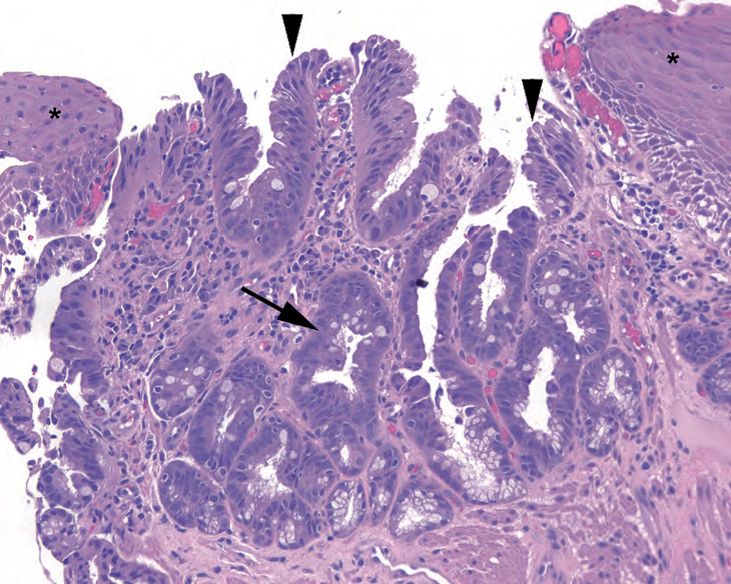
Figure 6.5. Low-grade dysplasia in Barrett’s esophagus. The cells begin to lose polarity and organization, with nuclei becoming more pleomorphic and lifting up off the basement membrane (arrow). The changes must extend to the surface (arrowheads) to count. Compare with the nondysplastic epithelium shown in Figure 6.4. Adjacent squamous epithelium can be seen on either side (asterisks).
图6.5.Barrett食管低级别异型增生。细胞开始失去极性和规则性,细胞核变得更加多形性,并从基底膜上抬起(箭头)。这些改变必须延伸到表面(箭头)才能当成低级别异型增生。与图6.4所示的非异型增生上皮相比较。两侧可见相邻鳞状上皮(星号)。
Progression to high-grade dysplasia (Figure 6.6) includes increasing atypia (loss of polarity, nuclei that begin to look like boulders: large with irregular outlines), mitotic activity (although mitoses alone are not worrisome), and architectural dysplasia (glands that are crowded and complex: budding, branching, cribriform, papillary, or villiform). High-grade dysplasia tends to be diagnosed in situations in which you are worried about invasive carcinoma but cannot prove it. Think of high-grade dysplasia as synonymous with carcinoma in situ.
向高级别异型增生的进展(图6.6)包括细胞异型性增加(极性丧失,细胞核开始看起来像巨石:大,轮廓不规则)、核分裂活跃(仅有核分裂并不令人担忧)和结构异型性增加(腺体拥挤而复杂:出芽、分枝、筛状、乳头或绒毛状)。在担心浸润性癌但无法证明浸润的情况下,往往可以诊断为高级别异型增生。将高级别异型增生视为原位癌的同义词。
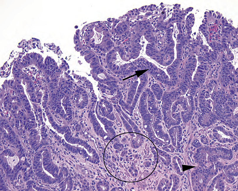
Figure 6.6. High-grade dysplasia. Nuclei here show marked hyperchromasia, pleomorphism, and disorganization (arrow), and the glands are beginning to show cribriform growth ( arrowhead). There is a focus suspicious for invasion highlighted in the circle.
图6.6.高级别异型增生。细胞核明显深染、多形性和紊乱(箭号),腺体开始显示筛状生长(箭头)。圆圈中突出显示了一处可疑浸润的病灶。
The next step along the malignancy progression is invasive carcinoma (Figure 6.7). As in other organs, clues to invasion include a ragged basement membrane border, single cells i nfiltrating, and a desmoplastic stromal response. Note that in the esophagus, unlike in the colon, intramucosal carcinoma (invasive adenocarcinoma confined to the lamina propria) is thought to have metastatic potential and thus is considered a “T1” lesion (not “Tis”) in the TNM (tumor, node, metastasis) staging classification. This is due to the presence of lymphatics in the lamina propria of the esophagus.
沿着恶性进展途径的下一步是浸润性癌(图6.7)。与其他器官一样,浸润的线索包括基底膜边缘破损、单个细胞浸润和促结缔组织增生性间质反应。注意食管与结肠不同,食管粘膜内癌(局限于固有层的浸润性腺癌)视为具有转移潜能,因此在TNM(肿瘤、淋巴结、转移)分期分类中视为是“T1”病变(而不是“Tis”)。这是由于食管固有层中存在淋巴管。
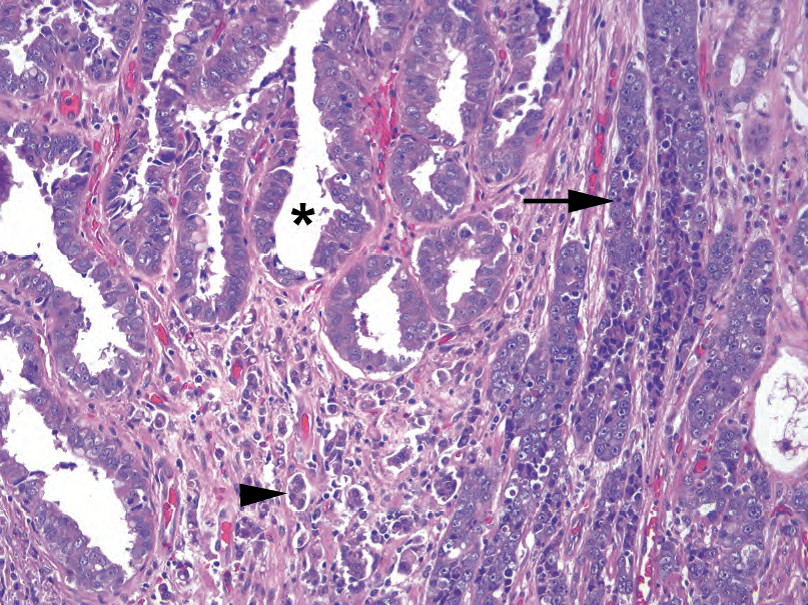
Figure 6.7. Invasive adenocarcinoma. The carcinoma can be seen invading the stroma as glands (asterisk), cords (arrow), and single cells (arrowhead). Notice the prominent nucleoli, which are not typically a feature of dysplasia.
图6.7.浸润性腺癌。癌细胞以腺体(星号)、条索(箭头)和单细胞(箭头)的形式侵入间质。注意显著的核仁,核仁通常不是异型增生的特征。
鳞状上皮异型增生(Squamous Dysplasia)
Within the squamous epithelium, dysplasia, carcinoma in situ, and invasive squamous cell carcinoma are diagnosed by criteria similar to those for the cervix. Dysplastic changes include enlarged, pleomorphic nuclei, increased nuclear/cytoplasmic ratio (a general blueness at low power), mitoses above the basal layer, and loss of order and polarity. Prominent nucleoli are more consistent with reactive/reparative changes. Dysplasia begins at the base and progresses to the surface. If the changes persist all the way to the surface, it is carcinoma in situ. Invasion can be hard to identify; a deep pushing front is not necessarily invasion. Look for deep aberrant keratinization (pinking up) and single cells trailing off as a clue to invasive carcinoma.
在鳞状上皮内,异型增生、原位癌和浸润性鳞状细胞癌的诊断标准与宫颈相似。异型增生改变包括核增大、多形性、核/质比增加(低倍镜下总体上呈蓝色)、基底层上方核分裂、失去规则性和极性。显著的核仁更符合反应性/修复性改变。异型增生从基底部开始,向表面延伸。如果这种改变一直持续到表面,那就是原位癌。浸润可能很难识别;深部推挤性前沿不一定是浸润。寻找深层异常角化(变红)和单个细胞脱落作为浸润性癌的线索。
息肉(Polyps)
A reasonable differential for a polypoid structure in the esophagus includes the following:
食管息肉样结构的合理鉴别诊断包括:
良性(Benign)
° Inflammatory fibroid polyp: vascular, inflamed, fibrous stroma covered by benign squamous epithelium, may be ulcerated; looks like granulation tissue
° 炎性纤维性息肉:良性鳞状上皮覆盖的血管、炎症、纤维性间质,可能有溃疡;看起来像肉芽组织
° Fibrovascular polyp: fibrovascular core covered by benign squamous epithelium, plus or minus fat
° 纤维血管性息肉:良性鳞状上皮覆盖的纤维血管轴心,伴或不伴脂肪
° Squamous papilloma: fibrovascular core covered by hyperplastic, but benign, squamous epithelium
° 鳞状上皮乳头状瘤:增生的但良性的鳞状上皮覆盖的纤维血管核心
° Submucosal nodules such as leiomyoma and granular cell tumor
° 粘膜下结节,如平滑肌瘤和颗粒细胞瘤
恶性(Malignant)
° Verrucous squamous cell carcinoma
° 疣状鳞状细胞癌
° Other carcinomas
° 其他癌
食管肿瘤(Neoplasms in the Esophagus)
One way to create a differential of neoplasms within an organ is to list all of the normal cell types and then think about what tumors can arise from each one. In the esophagus, the whopping majority of cancers arise from the epithelium and therefore are squamous or adenocarcinoma, but soft tissue tumors, although unusual, can occur. These include leiomyoma, granular cell tumor, hemangioma, angiosarcoma, and others.
在一个器官内,形成肿瘤的鉴别诊断的一种方法是列出所有的正常细胞类型,然后考虑每种细胞类型会产生什么肿瘤。在食道中,绝大多数癌症来自上皮,因此是鳞癌或腺癌。软组织肿瘤虽然少见,但也可能发生,包括平滑肌瘤、颗粒细胞瘤、血管瘤、血管肉瘤,等。
来源:
The Practice of Surgical Pathology:A Beginner’s Guide to the Diagnostic Process
外科病理学实践:诊断过程的初学者指南
Diana Weedman Molavi, MD, PhD
Sinai Hospital, Baltimore, Maryland
ISBN: 978-0-387-74485-8 e-ISBN: 978-0-387-74486-5
Library of Congress Control Number: 2007932936
© 2008 Springer Science+Business Media, LLC
仅供学习交流,不得用于其他任何途径。如有侵权,请联系删除。
本站欢迎原创文章投稿,来稿一经采用稿酬从优,投稿邮箱tougao@ipathology.com.cn
相关阅读
 数据加载中
数据加载中
我要评论

热点导读
-

淋巴瘤诊断中CD30检测那些事(五)
强子 华夏病理2022-06-02 -

【以例学病】肺结节状淋巴组织增生
华夏病理 华夏病理2022-05-31 -

这不是演习-一例穿刺活检的艰难诊断路
强子 华夏病理2022-05-26 -

黏液性血性胸水一例技术处理及诊断经验分享
华夏病理 华夏病理2022-05-25 -

中老年女性,怎么突发喘气困难?低度恶性纤维/肌纤维母细胞性肉瘤一例
华夏病理 华夏病理2022-05-07


共0条评论