外科病理学实践:诊断过程的初学者指南 | 第5章 意义有限的杂类标本
第5章 意义有限的杂类标本
(译注,原文标题是Ditzels,迪泽尔(俚语)。根据下文,意思可能是一些常见标本,临床送检主要目的不是为了诊断,而是一种记录。因为按规范,所以离体器官组织都要送病理。所以它们的临床意义和教学意义都是有限的。如果硬要按字面意思翻译,可能相当于宋元小说中的“那话儿”。一笑,哈哈)
Ditzel (slang): A word used to describe any part of the body that is not ordinarily appropriate for everyday conversation. “Susan is always walking down the hall with her ditzels hanging out.” (http://www.urbandictionary.com/)
迪泽尔(俚语):一个用来描述身体中通常不适合日常谈话的任何部位的词。“苏珊总是在大厅里闲逛,她的小弟弟们总是挂在外面。”
Ditzels are small specimens with limited educational potential. For the purposes of this chapter, these are all specimens with no suspicion or history of malignancy. They often have about three possible diagnoses and a reduced billing charge because of their limited complexity. Until you get experience with them, they slow you down inordinately at the grossing bench and at the microscope as you struggle to get the “right” wording and obsess over whether what you see is pathologic or normal. After all, it is really embarrassing to get a ditzel wrong. What follows is a list of typical features, things not to miss, and a suggested wording for unremarkable specimens. However, diagnosis style may vary across institutions, so take your cues from your own attendings.
Ditzel是教学价值有限的小标本。在本章中,这些标本均无可疑或恶性病史。由于复杂性有限,他们通常有大约三种可能的诊断,并减少了患者的医疗费用。直到你对它们有了处理经验,在此之前,你会在取材板凳上和在显微镜下犹豫不决,浪费时间,努力获得“正确”的措词,纠结于你所看到的是病理性的还是正常的。毕竟,在这种标本上犯错误真的很尴尬。下面是一个典型特征的列表,不要错过的东西,以及对无特殊标本的建议措辞。然而,不同医疗机构的诊断方式可能有所不同,所以请从你自己的主治医生那里获得提示。
Cholesteatoma (Middle Ear)
胆脂瘤(中耳)
Grossing: A small, whole specimen is usually submitted.
大体检查:通常送检一件完整的小标本。
Histology: A cyst is formed by keratinizing epithelium and filled with flaky keratin. Other features can include inflammation, cholesterol clefts, and foreign body giant cells (Figure 5.1).
组织学:囊肿由角化上皮形成,充满片状角化物。其他特征包括炎症、胆固醇裂缝和异物巨细胞(图5.1)。
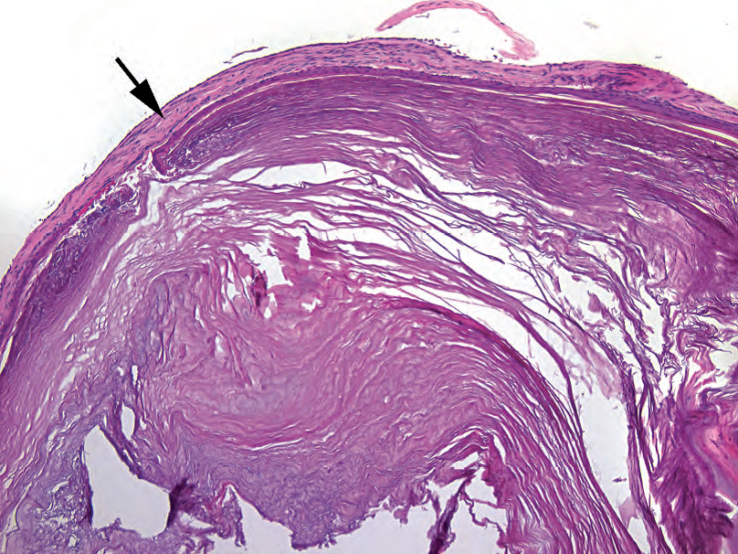
Figure 5.1. Cholesteatoma. The specimen is dominated by layers of pink keratin; the thin epithelium can be seen surrounding the keratin nodule (arrow).
图5.1.胆脂瘤。标本主要是一层粉红色角化物;角质结节周围可见薄层上皮(箭头)。
Rule out: Differential diagnosis of a middle ear mass includes inflammatory polyp, paraganglioma, middle ear adenoma, meningioma, and schwannoma.
排除:中耳肿块的鉴别诊断包括炎性息肉、副神经节瘤、中耳腺瘤、脑膜瘤和神经鞘瘤。
Sample sign out: Left middle ear (excision): Cholesteatoma or Fragments of keratinaceous debris (clinical cholesteatoma).
标本签发:左中耳(切除):胆脂瘤,或角化物碎片(临床胆脂瘤)
(译注:本人签发示例:
(左中耳切除标本)胆脂瘤。
或
(左中耳切除标本)送检角化物碎片,结合临床符合胆脂瘤。)
鼻窦内容物(Sinus Contents)
Grossing: Aspirate sent in a nylon bag. Submit one to two blocks, depending on volume. Use biopsy bags in cassettes.
大体检查:吸除标本装在尼龙袋中送检。根据体积,取材一到两个组织块,装入活检袋再放进包埋盒)。
(译注:国内多取纱布或滤纸代替活检袋)
Histology: Normal components include fragments of bone, respiratory and squamous mucosa, and mucous glands.
组织学:正常成分包括骨碎片、呼吸道粘膜和鳞状上皮粘膜以及粘液腺。
Chronic sinusitis: edema, acute and chronic inflammation
慢性鼻窦炎:水肿、急性和慢性炎症
Allergic fungal sinusitis: sheets of allergic mucin (Figure 5.2) and Charcot-Leyden crystals
过敏性真菌性鼻窦炎:成片的过敏性粘液(图5.2)和Charcot-Leyden晶体
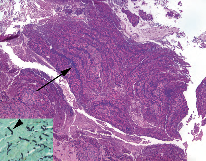
Figure 5.2. Allergic mucin in sinusitis. The allergic mucin takes on a characteristic tiger-striped appearance (arrow) as layers of eosinophils, mucin, and cell debris accumulate. Inset: A Gomori’s methenamine silver stain shows black fungal hyphae (arrowhead).
图5.2.鼻窦炎中的过敏性粘液。过敏性粘液呈现特征性虎斑外观(箭号),由数层嗜酸性粒细胞、粘液和细胞碎片堆积而成。插图:Gomori六胺银染色显示黑色真菌菌丝(箭头)。
Rule out: If allergic mucin is present, get a periodic-acid Schiff (PAS) or Gomori’s methenamine silver (GMS) stain to rule out fungus; other sinus lesions include polyps, papillomas, and unusual tumors.
排除:如果存在过敏性粘液,用PAS染色或六胺银(GMS)染色排除真菌;其他鼻窦病变包括息肉、乳头状瘤和少见肿瘤。
Sample sign out: Right and left sinus contents (aspiration): Chronic sinusitis or Fragments of respiratory mucosa with chronic inflammation or Allergic fungal sinusitis (a PAS stain highlights fungal organisms within the mucin).
标本签发:左右鼻窦内容物(吸除):慢性鼻窦炎,或呼吸道粘膜碎片伴慢性炎症,或过敏性真菌性鼻窦炎(PAS染色突出显示粘液内的真菌病原体)。
颈动脉和股动脉斑块(Carotid and Femoral Plaques)
Grossing: Specimen is essentially a cast of the artery lumen. Take one block of representative cross sections; this usually requires light decalcification.
大体检查:标本基本上是动脉腔的模子。取一块具有代表性的横截面;通常需要轻度脱钙。
Histology: The inner layer of the elastic arterial wall has a variable amount of atherosclerotic debris, calcification, and/or thrombus (Figure 5.3).
组织学:弹性动脉壁的内层有不同数量的动脉粥样硬化碎片、钙化和/或血栓(图5.3)。
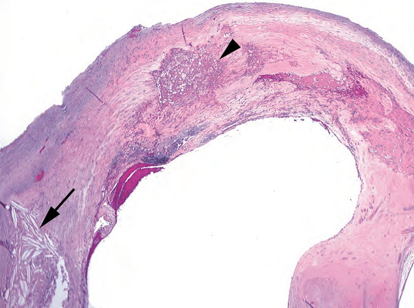
Figure 5.3. Carotid plaque. This represents the intimal surface of the artery, in which there may be calcification, foamy macrophages (arrowhead), cholesterol clefts (arrow), or inflammatory debris.
图5.3.颈动脉斑块。这代表动脉内表面,其中可能有钙化、泡沫状巨噬细胞(箭头)、胆固醇裂缝(箭号)或炎症碎片。
Sample sign out: Carotid artery, right (endarterectomy): Calcified atherosclerotic plaque.
标本签发:右侧颈动脉(动脉内膜切除术):钙化的动脉粥样硬化斑块。
椎间盘(Intervertebral Disc)
Grossing: Submit one block of representative or total material.
大体检查:送检一块代表性材料或全部材料。
Histology: Fibrocartilage and pulpy myxoid gel (the nucleus pulposus), possibly with fragments of bone (Figure 5.4), are present.
组织学:纤维软骨和髓样黏液凝胶(髓核),可能有骨碎片(图5.4)。
Sample sign out: Cervical disc (excision): Fragments of disc material.
标本签发:颈椎间盘(切除):破碎的椎间盘组织。
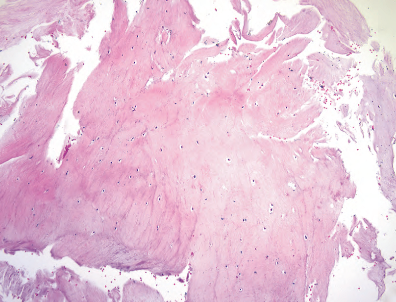
Figure 5.4. Intervertebral disc. The disc substance is paucicellular, with a homogeneous translucent stroma (ranging from myxoid to collagenous).
图5.4.椎间盘。椎间盘物质中细胞稀少,具有均匀的半透明间质(从粘液样到胶原化)。
胸腺(Thymus)
Grossing: Tissue may be from incidental thymectomy (heart surgeries), biopsy, or something else (such as parathyroid). Weigh it. Weight is a criterion for true thymic hyperplasia. Submit in total for small specimens or representative for thymectomy.
大体检查:组织可能来自偶然的胸腺切除术(心脏手术)、活检或其他情况(如甲状旁腺)。称重。重量是判断真性胸腺增生的标准。对于小标本,全部取材,而胸腺切除术标本选取代表性组织块。
Histology: Architecture is lobular with dark outer cortex and pale medulla (Figure 5.5). H assall’s corpuscles look like squamous nests. Germinal centers are not normal. Fatty replacement with age is normal.
组织学:结构为小叶状,外层为深染的皮质,中央为淡染的髓质(图5.5)。胸腺小体看似鳞状细胞巢。生发中心不是正常表现。随着年龄的增长,脂肪替代胸腺组织是正常的。
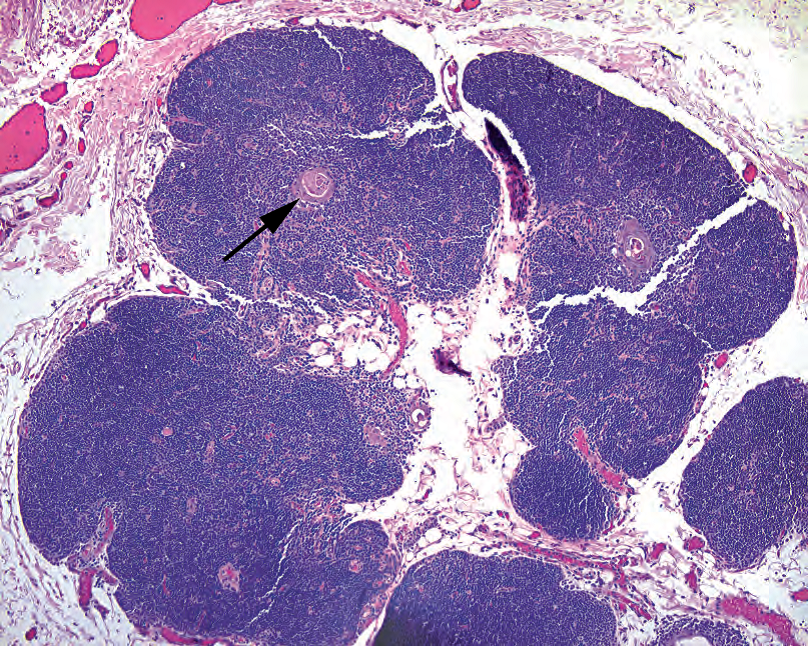
Figure 5.5. Thymus. The lobulated thymus resembles lymph node at low power but should not have germinal centers. Hassall’s corpuscles (arrow) are visible as pink whorls.
图5.5.胸腺。分叶状胸腺在低倍镜下类似淋巴结,但不应有生发中心。可见胸腺小体(箭头),表现为粉红色旋涡状。
Rule out: Distinguish from sheets of cells or obliterated architecture (thymoma).
排除:区别于成片细胞或结构消失(胸腺瘤)。
Sample sign out: Thymus (thymectomy) or “left inferior parathyroid” (excision): Histologically unremarkable thymus.
标本签发:胸腺(胸腺切除术)或“左下甲状旁腺”(切除术):组织学上无特殊的胸腺组织。
甲状旁腺(Parathyroid)
Grossing: Weigh it. Submit it in its entirety.
大体检查:称重。全部取材。
Histology: Features include monotonous round neuroendocrine cells with clear cytoplasm (chief cells) or abundant pink cytoplasm (oxyphil cells). Normal weight is around 50 mg; adenomas are usually >300 mg. Adenoma is a clinical diagnosis requiring evidence of normalized parathyroid hormone level after surgery. Hyperplasia and adenoma may look the same on the slide (Figure 5.6).
组织学:特征包括形态单一的圆形神经内分泌细胞伴透明细胞质(主细胞)或丰富粉红色细胞质(嗜酸性细胞)。正常重量约为50毫克;腺瘤通常>300毫克。腺瘤是一种临床诊断,需要手术后甲状旁腺激素水平恢复正常的证据。增生和腺瘤在切片上看起来可能相同(图5.6)。
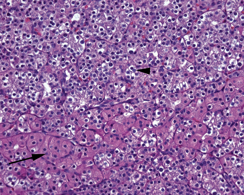
Figure 5.6. Parathyroid tissue. Normal parathyroid has two cell populations, the chief cells (arrowhead) and oxyphil cells (arrow).
图5.6.甲状旁腺组织。正常甲状旁腺有两个细胞群,主细胞(箭头)和嗜酸性细胞(箭号)。
Rule out: Carcinoma is very rare, but dense fibrotic bands and nuclear atypia are suggestive.The diagnosis of carcinoma requires capsular or vascular invasion.
排除:癌非常罕见,但致密的纤维束和核异型性提示癌。诊断癌需要包膜浸润或血管浸润。
Sample sign out: Left superior parathyroid (excision):Cellular parathyroid tissue (250 mg).
标本签发:左上甲状旁腺(切除):细胞丰富的甲状旁腺组织(250mg)。
心瓣膜(Heart Valves)
Grossing: There is much information to be gained in grossing. Review your grossing manual for details. Note the presence of vegetations, commissural fusion, calcification, and redundancy.
大体检查:大体检查可以获得很多信息。查看你的取材手册以了解详细信息。注意赘生物、连合融合、钙化和冗余的存在。
Histology: Look for myxoid degeneration, calcification, and adherent vegetations (Figure 5.7).
组织学:查找粘液样变性、钙化和黏附的赘生物(图5.7)。
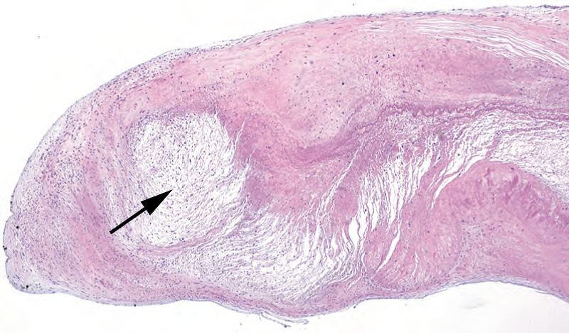
Figure 5.7. Myxoid degeneration, heart valve. In the free end of this heart valve, there is an attenuated pale area of myxoid degeneration (arrow). Calcifications and vegetations may also be seen.
图5.7.黏液样变性,心瓣膜。在这个心瓣膜的游离端,有一个粘液样变性的淡染区域(箭头)。也可以看到钙化和赘生物。
Rule out: Use Gram Weigert and GMS stains on vegetations to rule out bacteria or fungus.
排除:对赘生物使用Gram Weigert染色和GMS染色,排除细菌或真菌。
Sample sign out: Aortic valve (excision): Valve with myxoid degeneration and calcification or Valve with adherent fibrinopurulent debris. Numerous Gram-positive cocci are seen on Gram Weigert stain.
标本签发:主动脉瓣(切除):瓣膜伴黏液样变性和钙化,或瓣膜伴粘附的纤维素碎屑。Gram Weigert染色可见大量革兰阳性球菌。
胆囊(Gallbladder)
Grossing: The first section should be a cross section of the cystic duct margin. Open the gallbladder, look for stones, and take two sections of the wall. Note the wall thickness and describe the mucosa. All three fragments can go in one cassette.
大体检查:取材第一个组织块应该是胆囊管切缘的横切面。打开胆囊,查找结石,取两块胆囊壁。记录胆囊壁厚度并描述粘膜。所有三个组织块都可以放在一个包埋盒中。
Histology: The gallbladder is lined by a single layer of columnar epithelium in folds, overlying a fibromuscular layer that sometimes contains Rokitansky-Aschoff sinuses (infolded mucosa) or ducts of Luschka. Cholecystitis can range from mild lymphoplasmacytic inflammation to transmural acute inflammation. Cholesterolosis is the accumulation of foamy macrophages (Figure 5.8).
组织学:胆囊被覆一层有皱襞的柱状上皮,下方是一层纤维肌层,有时含有Rokitansky-Aschoff窦(皱褶粘膜)或Luschka导管。胆囊炎的程度范围从轻微的淋巴浆细胞炎症到跨壁急性炎症。胆固醇贮积病是泡沫状巨噬细胞的积聚(图5.8)。
(译注:胆囊没有粘膜肌层;胆固醇贮积病临床上又称为胆固醇息肉)
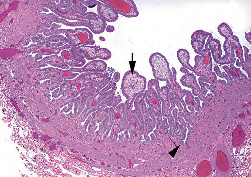
Figure 5.8. Gallbladder with cholesterolosis. The mucosal folds are distended with foamy macrophages (arrow), called cholesterolosis. Inflammation is minimal in this example. Rokitansky-Aschoff sinuses can penetrate deeply into the gallbladder wall (arrowhead).
图5.8.胆囊伴胆固醇贮积病。粘膜皱襞因泡沫状巨噬细胞(箭号)而膨胀,称为胆固醇贮积病。本例炎症轻微。Rokitansky-Aschoff窦可深入胆囊壁(箭头)。
Rule out: Dysplasia or carcinoma is rare in an isolated cholecystectomy.
排除:异型增生或癌在孤立性胆囊切除术中很少见。
Sample sign out: Gallbladder (cholecystectomy): Chronic cholecystitis, cholelithiasis, and cholesterolosis or Acute and chronic cholecystitis.
标本签发:胆囊(胆囊切除术):慢性胆囊炎、胆石症和胆固醇贮积病,或急慢性胆囊炎。
阑尾(Appendix)
Grossing: The first section should be a cross section of the proximal margin, inked or otherwise marked as margin. Then cut off the tip and take a longitudinal section (U shaped). Breadloaf the remainder, and take one to two cross sections. Look for nodules, fecaliths, hemorrhage, and pus.
加粗:取材第一个组织块应该是近端边缘的横切面,用墨水或其他方式标记为边缘。然后切断尖端并取一个纵切面(U形)。剩余组织切成面包片,取一到两个横截面。查找结节、粪便、出血和脓液。
Histology: Normal histology is a colonic mucosa with abundant lymphoid aggregates. Chronic inflammation is not significant, but neutrophils are, whether in the mucosa, wall (transmural inflammation; Figure 5.9), or serosa (serositis). Serositis without transmural inflammation suggests another abdominal source.
组织学:正常组织学为结肠粘膜,有丰富的淋巴组织聚集。慢性炎症不明显,但中性粒细胞明显,无论是粘膜、阑尾壁(跨壁炎症;图5.9)还是浆膜(浆膜炎)。无跨壁炎症的浆膜炎提示腹腔其他部位的炎症来源。
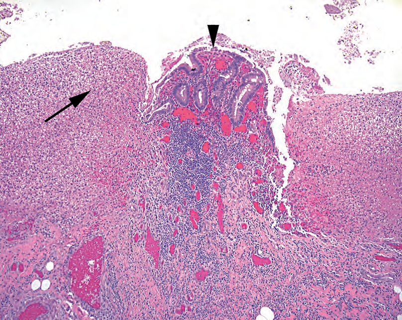
Figure 5.9. Appendicitis. In this close-up view, a small amount of residual colonic-type mucosa is visible (arrowhead), surrounded by mounds of fibrinopurulent debris (arrow).
图5.9.阑尾炎。在这张特写图中,可见少量残留的结肠型粘膜(箭头),周围是成堆的纤维素性碎屑(箭号)。
Rule out: Carcinoid in the tip and pools of mucin in the wall (as in a mucinous cystadenoma or carcinoma) should be ruled out.
排除:应排除阑尾尖端的类癌和阑尾壁上的粘液池(如粘液性囊腺瘤或癌中所见)。
Sample sign out: Appendix (appendectomy): Acute transmural appendicitis with serositis or Histologically unremarkable appendix.
标本签发:阑尾(阑尾切除术):急性跨壁阑尾炎伴浆膜炎,或组织学上无特殊的阑尾。
Hernia Sac
疝囊
Grossing: Submit a representative section.
大体检查:选取代表性组织块。
Histology: A pouch of fibroadipose tissue is lined with mesothelium, which can be reactive or proliferative (Figure 5.10).
组织学:被覆间皮的囊袋状纤维脂肪组织,间皮可呈反应性或增殖性(图5.10)。
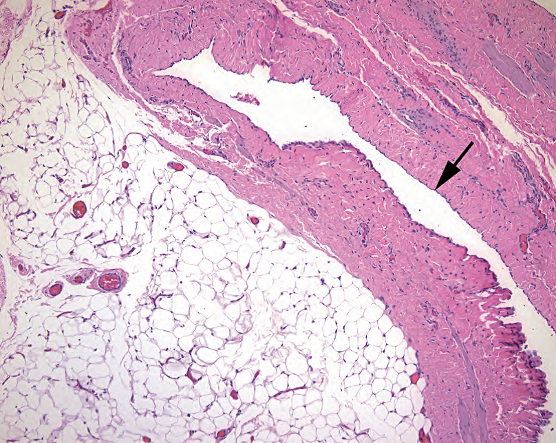
Figure 5.10. Hernia sac. Thick fibrous tissue and fat characterize the typical hernia sac. In this section, the delicate mesothelial lining (arrow) is visible.
图5.10.疝囊。厚的纤维组织和脂肪是典型疝囊的特征。这部分可以看到菲薄的被覆间皮(箭头)。
Rule out: A piece of the vas deferens (warrants an immediate call to the surgeon), incarcerated bowel, and metastatic tumor (especially incisional hernias) should be ruled out.
排除:应该排除输精管(需要立即呼叫外科医生)、肠嵌顿和转移性肿瘤(尤其是切口疝)的可能性。
Sample sign out: Soft tissue, right inguinal (herniorrhaphy): Hernia sac or Fibrovascular tissue (clinical hernia sac).
标本签发:右侧腹股沟软组织(疝修补术):疝囊,或纤维血管组织(临床疝囊)。
瘢痕修复(Scar Revision)
Grossing: Breadloaf and take representative sections through the scar. Note that this does not apply to reexcision of a skin cancer or melanoma.
大体检查:切成面包片,通过瘢痕选取代表性组织块。请注意,这种取材方法不适用于皮肤癌或黑色素瘤的再次切除。
Histology: Dermal scar has dense fine pink collagen, no appendages, and thin epithelium (Figure 5.11). Recent injury or surgery will show hemorrhage, foamy macrophages, inflammation and granulation tissue, suture material, and foreign body–type giant cells.
组织学:皮肤瘢痕有致密的细腻的粉红色胶原,无皮肤附属器,上皮薄(图5.11)。近期的损伤或手术将显示出血、泡沫状巨噬细胞、炎症和肉芽组织、缝合材料和异物型巨细胞。
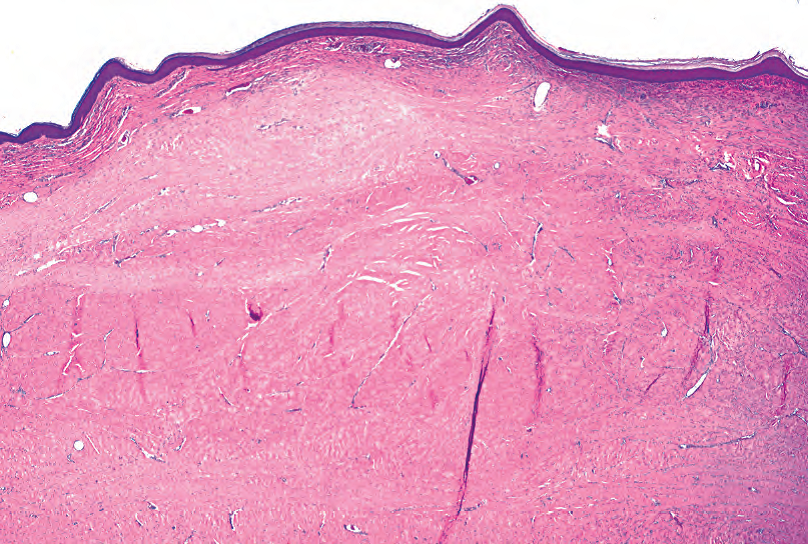
Figure 5.11. Dermal scar. Pale and homogeneous collagen underneath the epidermis, with obliteration of adnexal structures, is typical of scar formation.
图5.11.皮肤瘢痕。表皮下均匀淡染的胶原,伴附属器结构的闭塞,是典型的瘢痕形成。
Rule out: Exclude tumor, if there is a history of tumor, and abscess.
排除:如果有肿瘤病史,排除肿瘤。并排除脓肿
Sample sign out: Left abdominal wall (scar revision): Skin with dermal scar, negative for tumor or Skin with biopsy site changes and suture material.
标本签发:左腹壁(瘢痕修复):皮肤伴皮肤瘢痕,未见肿瘤,或皮肤伴活检部位改变和缝合材料。
股骨头或肱骨头、膝盖骨(Femoral or Humeral Head, Knee Bones)
Grossing: Use a bone saw to cross section the bone and get a 2-mm slice. Describe eburnation (absence of cartilage), osteophytes, femoral neck (fracture vs. surgical), infarcts, and subchondral cysts. Sample the articular surface, plus the margin or fracture site in fracture cases. Submit for routine decalcification.
大体检查:用骨锯横切骨骼,取一块2毫米厚的组织块。描述磨损(软骨缺失)、骨赘、股骨颈(骨折对比手术)、梗死和软骨下囊肿。取关节面和切缘,或骨折病例的骨折部位。送检常规脱钙。
Histology: Healthy bone has a thick cartilage layer with a smooth surface and marrow between the trabeculae. Look for the following:
组织学:健康骨骼有一层厚的软骨层,表面光滑,小梁之间有骨髓。查看以下内容:
Osteoarthritis: uneven and ragged or absent cartilage, clonal nests of chondrocytes, dual tide-line, thickened and sclerotic subchondral bone, subchondral cysts, fibrosis, and granulation tissue within marrow (Figure 5.12)
骨关节炎:软骨不均匀、参差不齐或缺失,软骨细胞克隆巢(同源细胞巢),双潮线,软骨下骨增厚和硬化,软骨下囊肿,纤维化,骨髓内肉芽组织(图5.12)
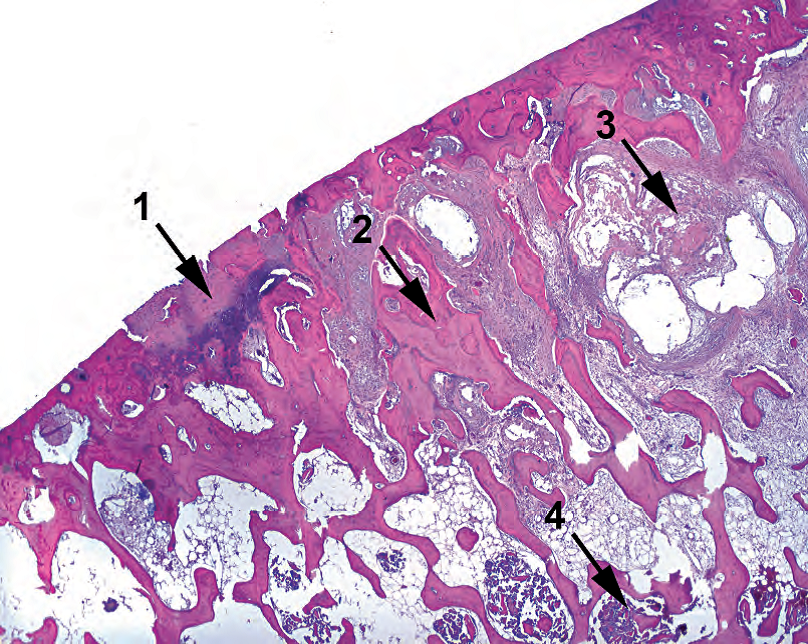
Figure 5.12. Osteoarthritis. Features include (1) eroded cartilage, in this case nearly absent, and irregular mineralization of the cartilage, seen here as a dark purple stain; (2) thickening of the subchondral bony trabeculae; (3) myxoid degeneration of the subchondral bone, forming cyst-like spaces; and (4) some residual hematopoietic marrow.
图5.12.骨关节炎。特征包括:(1)软骨磨损,在本例中几乎不存在,软骨不规则矿化,此处表现为深紫色斑点;(2)软骨下骨小梁增厚;(3)软骨下骨粘液样变性,形成囊肿样间隙;(4)部分残存的造血性骨髓。
Osteonecrosis: loss of basophilia and nuclei in the marrow, fat cells, and osteocytes; fat necrosis; hemorrhage (Figure 5.13)
骨坏死:骨髓、脂肪细胞和骨细胞失去嗜碱性和细胞核;脂肪坏死;出血(图5.13)
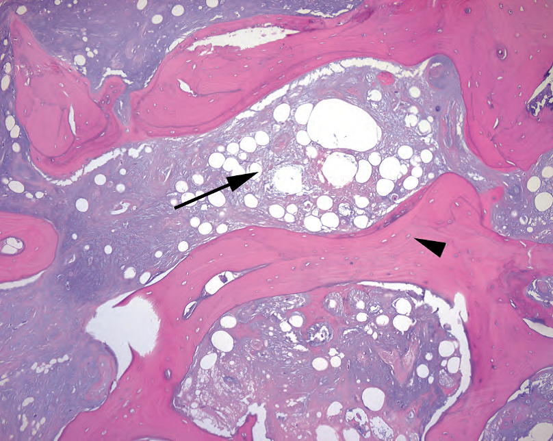
Figure 5.13. Osteonecrosis. The necrotic marrow is the most eye-catching feature (arrow), showing fat necrosis and an absence of marrow elements. On close examination, the osteocytes within lacunae are also dead or missing (arrowhead).
图5.13.骨坏死。坏死的骨髓是最引人注目的特征(箭号),显示脂肪坏死和骨髓成分缺失。仔细检查,骨陷窝内的骨细胞也死亡或缺失(箭头)。
Osteopenia: markedly thinned trabeculae
骨质减少:骨小梁明显变薄
Metastatic tumor: out-of-place cells in the area of fracture
转移性肿瘤:骨折区域的不适当细胞
Sample sign out: Right femoral head (arthroplasty): Femoral head with osteonecrosis and fracture or Bone and cartilage with degenerative changes or Osteoarthritis
标本签发:右股骨头(关节成形术):股骨头伴骨坏死和骨折,或骨和软骨伴退行性改变,或骨关节炎。
截肢(Amputated Limbs)
Grossing: It is gross, all right. Document the extent of gangrene, ulcers, venous stasis, trauma, and so forth, as well as level of amputation and the viability of the margin. In vascular or infectious disease, section the vascular margin (i.e., popliteal). Take representative sections from the worst area (soft tissue) and margin. Tissue from the bony margin is usually not necessary.
大体检查:真恶心,好吧。记录坏疽、溃疡、静脉淤滞、创伤等的程度,以及截肢水平和边缘是否存活。在血管病或感染性疾病中,切取血管切缘。从最差区域(软组织)和边缘选取代表性组织块。骨边缘的组织通常是不必要的。
Histology: Look for gangrenous necrosis (Figure 5.14), ulceration, scar, granulation tissue, and inflammation. Evaluate vessel for atherosclerotic disease.
组织学:检查坏疽性坏死(图5.14)、溃疡、瘢痕、肉芽组织和炎症。评估动脉粥样硬化疾病。
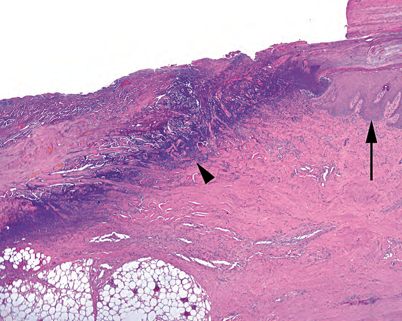
Figure 5.14. Gangrene. In this gangrenous ulcer of the toe, the epidermis is visible to the right (arrow), while the ulcer bed to the left shows an obliteration of epidermis and dermis, with a dense blue line of debris representing dying bacteria and cells (arrowhead).
图5.14.坏疽。在脚趾坏疽性溃疡中,右侧可见表皮(箭号),而左侧溃疡床显示表皮和真皮闭塞,有一条密集的蓝色碎片线,代表死亡的细菌和细胞(箭头)。
Rule out: Invasive fungal disease in a neutropenic patient (requires more extensive sampling of the margin) should be ruled out.
排除:中性粒细胞减少症患者的侵袭性真菌病(需要更广泛的边缘取样)。
Sample sign out: Left foot (amputation): Foot with gangrenous necrosis. Atherosclerotic vessels are identified. Surgical margin appears viable.
标本签发:左脚(截肢):足伴坏疽性坏死。可见动脉粥样硬化血管。手术切缘似乎是存活的。
脂肪瘤(Lipoma)
Grossing: Measure it. It never hurts to ink it. Submit thin slices (one per centimeter), and give them a nice long fixation time. Sample areas that are fibrous, fleshy, hemorrhagic, or otherwise nonfatty.
大体检查:测量。墨水无法染色。切取薄片(每厘米一片),要很长的固定时间。纤维状、肉样、出血性或其他非脂肪性区域都要取材。
Histology: The definition of a lipoma is a neoplasm of mature fat cells. Fibrous tissue is okay. In fact, there are at least eight benign varieties of lipoma (fibrolipoma, myxolipoma, chondroid lipoma, myolipoma, myelolipoma, spindle cell lipoma, pleomorphic lipoma, and angiolipoma), all fat with something extra.
组织学:脂肪瘤的定义是成熟脂肪细胞的肿瘤。纤维组织没问题。事实上,至少有八种良性脂肪瘤(纤维脂肪瘤、粘液脂肪瘤、软骨样脂肪瘤、平滑肌脂肪瘤、髓脂肪瘤、梭形细胞脂肪瘤、多形性脂肪瘤和血管脂肪瘤),所有脂肪都含有额外的成分。
Rule out: Exclude liposarcoma. Clinical features that are suspicious include a large deep-seated circumscribed mass in the thigh, shoulder, retroperitoneum, or mesentery of an adult. Histologic features include chicken-wire vessels (a distinct lacy honeycomb network), atypical cells (large hyperchromatic nuclei), and lipoblasts (Figure 5.15). More details on liposarcomas are given in Chapter 28.
排除:排除脂肪肉瘤。可疑的临床特征包括成人大腿、肩部、腹膜后或肠系膜的大体积、位置深的局限性肿块。组织学特征包括鸡笼样血管(明显的花边蜂窝状网络)、非典型细胞(大的深染核)和脂肪母细胞(图5.15)。关于脂肪肉瘤的更多细节见第28章。
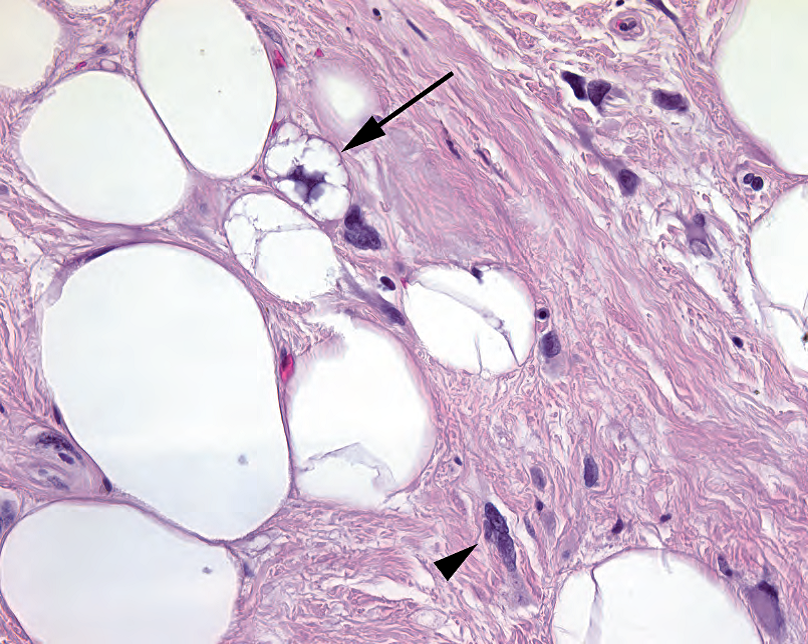
Figure 5.15. Lipoblast in an atypical lipoma. What you do not want to see in your lipoma—lipoblasts (arrow), with small fat vacuoles indenting the nucleus and atypical hyperchromatic cells within the fibrous stroma (arrowhead).
图5.15.非典型脂肪瘤中的脂肪母细胞。你不想在脂肪瘤中看到的—脂肪母细胞(箭号),小的脂肪空泡挤压细胞核,纤维间质内有非典型深染细胞(箭头)。
Sample sign out: Soft tissue, left flank (excision): Lipoma (12 cm) or Fibrovascular tissue and mature adipose tissue (clinical lipoma)
标本签发:左侧软组织(切除):脂肪瘤(12cm),或纤维血管组织和成熟脂肪组织(临床脂肪瘤)。
来源:
The Practice of Surgical Pathology:A Beginner’s Guide to the Diagnostic Process
外科病理学实践:诊断过程的初学者指南
Diana Weedman Molavi, MD, PhD
Sinai Hospital, Baltimore, Maryland
ISBN: 978-0-387-74485-8 e-ISBN: 978-0-387-74486-5
Library of Congress Control Number: 2007932936
© 2008 Springer Science+Business Media, LLC
仅供学习交流,不得用于其他任何途径。如有侵权,请联系删除。
本站欢迎原创文章投稿,来稿一经采用稿酬从优,投稿邮箱tougao@ipathology.com.cn
相关阅读
 数据加载中
数据加载中
我要评论

热点导读
-

淋巴瘤诊断中CD30检测那些事(五)
强子 华夏病理2022-06-02 -

【以例学病】肺结节状淋巴组织增生
华夏病理 华夏病理2022-05-31 -

这不是演习-一例穿刺活检的艰难诊断路
强子 华夏病理2022-05-26 -

黏液性血性胸水一例技术处理及诊断经验分享
华夏病理 华夏病理2022-05-25 -

中老年女性,怎么突发喘气困难?低度恶性纤维/肌纤维母细胞性肉瘤一例
华夏病理 华夏病理2022-05-07


共0条评论