外科病理学实践:诊断过程的初学者指南 | 第17章 子宫(Uterus)
第17章 子宫(Uterus)
The endometrium is a cycling glandular and stromal layer overlying the myometrium of the uterus. The appearance varies widely across different phases of the menstrual cycle, pregnancy, and menopause. Common reasons for performing an endometrial biopsy include the following:
子宫内膜覆盖在子宫肌层上方,是一种周期性腺体和间质层。在月经周期的不同阶段、怀孕和更年期,其形态学差异很大。子宫内膜活检的常见原因包括:
Abnormal vaginal bleeding
阴道异常出血
A “thickened endometrial stripe” found on ultrasound, suggesting hyperplasia or carcinoma
超声检查发现“子宫内膜增厚”,提示增生或癌
Part of an infertility workup
不孕症检查
Follow-up study for women with a history of hyperplasia who have been conservatively treated with hormones
对有增生病史且接受激素保守治疗的妇女的随访研究
子宫内膜活检的观察方法(The Approach to the endometrial biopsy)
The reason for the biopsy will influence your approach to the slide. Regardless of history, start by differentiating between atrophic, inactive, proliferative, secretory, and hormone-treated endometrium. On low power, survey the epithelium to get a feel for the glands and stroma:
活检原因会影响你观察切片的方法。不管病史如何,首先要区分萎缩性、非活动性、增殖性、分泌性和激素治疗的子宫内膜。在低倍镜下检查上皮,获得腺体和间质的一种感觉:
Atrophic endometrium has a low gland-to-stroma ratio, and the glands are thin, with an almost cuboidal epithelium, and no mitoses. In biopsy specimens, they tend to come off in thin strips that look like hair pins (Figure 17.1).
萎缩性子宫内膜:腺体与间质的比值低,腺体薄,几乎为立方上皮,无核分裂。在活检标本中,它们往往脱落成细条状,类似发夹(图17.1)。
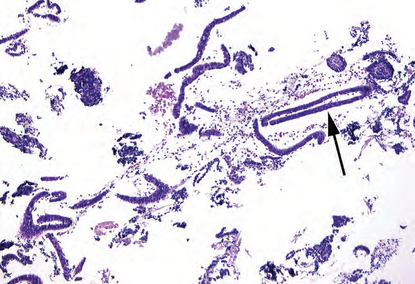
Figure 17.1. Atrophic endometrium. When curetted, the epithelium typically comes off in thin strips resembling hairpins (arrow). The specimen is also scant.
图17.1 萎缩性子宫内膜。刮除时,上皮通常脱落成细条状,类似发夹(箭)。样本量也很少。
Inactive to proliferative endometrium has a fuller, blue look to the stroma and a gland-tostroma ratio of about 1:1 in proliferative endometrium (less in inactive). The glands are simple tubular structures that stand out as dark blue “donuts” with pseudostratified nuclei (slight variation in nuclear location, but predominantly basal) and columnar epithelium (Figure 17.2). If mitoses are readily visible in the glands, the endometrium is proliferative. Absence of mitoses indicates an inactive endometrium.
非活动性到增殖性子宫内膜:间质更丰富,呈蓝色。增殖性子宫内膜中,腺体与子宫内膜的比值约为1:1(非活动性子宫内膜中比值更小)。腺体是简单的管状结构,呈深蓝色的“甜甜圈”形状,具有假复层细胞核(核位置略有变化,但主要位于基底)和柱状上皮(图17.2)。如果腺体中容易看到核分裂,则为增殖性子宫内膜。无核分裂提示非活动性子宫内膜。
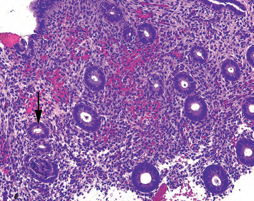
Figure 17.2. Proliferative endometrium. Multiple donut-shaped glands are visible, with dark oblong nuclei and frequent mitoses (arrow).
图17.2 增殖性子宫内膜。可见多个甜甜圈状腺体,细胞核深染,呈长方形,核分裂易见(箭)。
Secretory endometrium has prominent spiral arterioles and variably edematous stroma so that the stromal cells look almost like naked nuclei floating in water. The glands are notable for cytoplasmic secretory vacuoles and secretions in the lumen (Figure 17.3). Later secretory stroma begins to get decidualized, or acquires pink cytoplasm, and the glands lose their vacuoles and acquire low cuboidal pink cells, ragged luminal edges, and a tortuous spiral shape. You should not see mitoses in secretory glands.
分泌性子宫内膜:有显著的螺旋小动脉和不同程度的水肿性间质,因此间质细胞看起来就像是漂浮在水中的裸核。腺体具有明显的胞质分泌空泡和腺腔内分泌物(图17.3)。随后,分泌性间质开始蜕膜化,或获得粉红色细胞质,腺体失去分泌空泡,变成低立方形粉红色细胞,腺腔边缘参差不齐,呈扭曲的螺旋状。分泌性腺体不应看到核分裂。
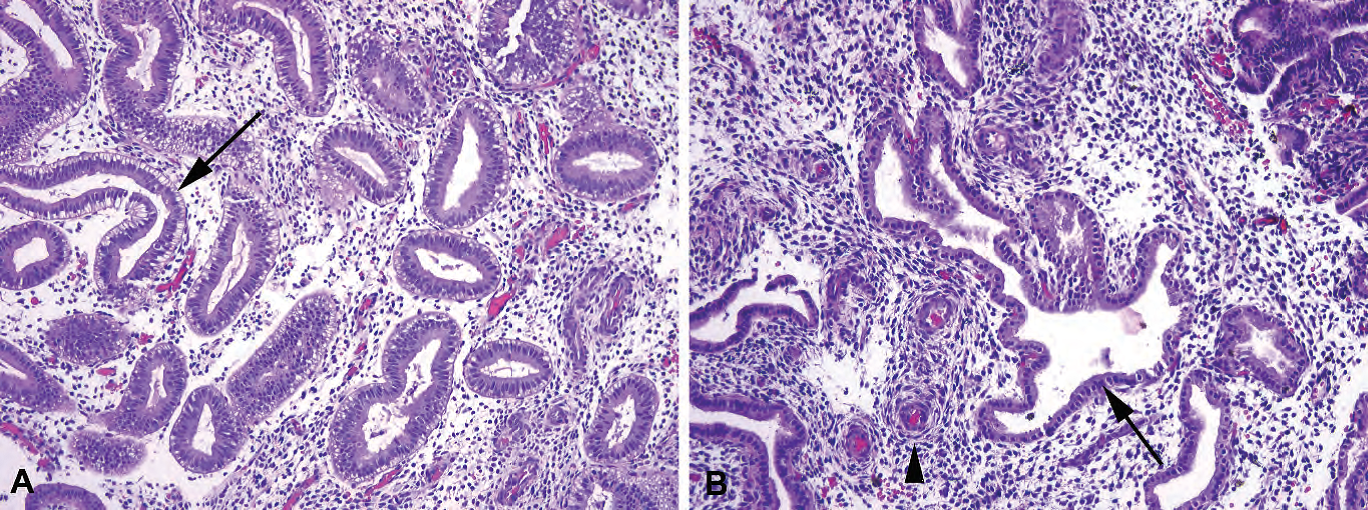
Figure 17.3. Secretory endometrium, various phases. (A) In early secretory endometrium, the glands have become tortuous in shape, and prominent cytoplasmic vacuoles are present (subnuclear, in this example; arrow). (B) Later in the secretory phase, the cytoplasmic vacuoles are gone, and the epithelium is more cuboidal in shape, with small round nuclei (arrow). The stroma is edematous, and early decidualization (accumulation of pink cytoplasm) is beginning around the spiral arteries (arrowhead).
图17.3 不同阶段的分泌性子宫内膜。(A)在早期分泌性子宫内膜中,腺体形状弯曲,出现显著的胞质空泡(本例为核下空泡;箭)。(B)在晚分泌期,胞质空泡消失,上皮变得更像立方形,细胞核小而圆(箭)。间质水肿,早期蜕膜化(粉红色细胞质的聚积)开始出现在螺旋动脉(箭头)周围。
Progestin-treated endometrium, like gestational endometrium, has a very decidualized stroma (plump pink cells with visible cytoplasm), but is paired with attenuated, flattened gland epithelium (Figure 17.4). These changes are due to the unopposed progesterone exposure. Unopposed estrogen, on the other hand, has a proliferative effect, and increases the chance of hyperplasia or carcinoma. (Tamoxifen acts as an estrogen agonist in the endometrium.)
孕激素治疗的子宫内膜:就像妊娠期子宫内膜,具有非常明显的蜕膜化间质(丰满的粉红色细胞,胞质丰富),对应的腺体上皮变薄变扁平(图17.4)。这些改变是由于无对抗性孕酮暴露所致。另,无对抗性雌激素具有促进增殖作用,并增加增生或癌的机会。(他莫昔芬对子宫内膜有雌激素激动剂的作用。)
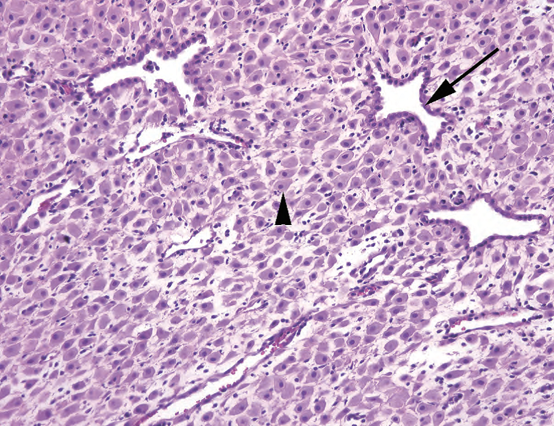
Figure 17.4. Progestin-treated endometrium. The glands are still tortuous in shape, like secretory endometrium, but the epithelium is markedly thinned (arrow). The stromal cells are decidualized (arrowhead), which means they have plump pink cytoplasm and distinct cell borders.
图17.4 孕激素治疗的子宫内膜。腺体形状仍然弯曲,像分泌性子宫内膜,但上皮明显变薄(箭)。间质细胞蜕膜化(箭头),即,具有丰满的粉红色细胞质和清晰的细胞边界。
Why are the endometrial characteristics important? Secretory endometrium, almost by definition, is not hyperplastic. Once you have established secretory change, you (usually) do not need to agonize over crowded glands. Because progesterone pushes the endometrium toward secretory change, it is used as treatment for hyperplasia; if you can prod the endometrium to complete the cycle and shed, the hyperplasia may go away.
为什么子宫内膜特征很重要?根据定义,分泌性子宫内膜几乎不是增生性。一旦你确定了分泌改变,你(通常)就不必为拥挤的腺体而苦恼了。由于孕酮促使子宫内膜发生分泌性改变,因此使用孕酮治疗增生;如果能刺激子宫内膜完成月经周期并脱落,增生可能会消失。
Next, within the biopsy fragments, look for possible causes of bleeding:
然后,在活检片段内,寻找出血的可能原因:
Benign endometrial polyp: Benign endometrial polyps are composed of fibrotic (pink and spindly) stroma, thick-walled vessels, and usually nonfunctional (atrophic) and/or cystically dilated glands (Figure 17.5).
良性子宫内膜息肉:纤维化(粉红色和梭形)间质、厚壁血管和通常无功能的(萎缩性)和/或囊性扩张的腺体(图17.5)。
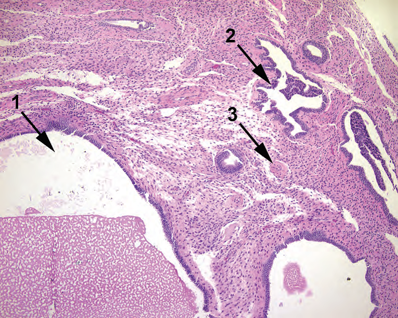
Figure 17.5. Benign endometrial polyp. This polyp shows cystic dilation of glands (1), secretory-type epithelium (2), and thickened arteries (3). The stroma is also pink, indicating a high collagen content.
图17.5 良性子宫内膜息肉。息肉显示腺体呈囊性扩张(1)、分泌型上皮(2)和厚壁动脉(3)。间质也是粉红色,提示胶原含量高。
Endometrial stromal breakdown: The stroma takes on a blurry blue look as it condenses into small dense aggregates (“blue balls”). The associated surface epithelium shows eosinophilic metaplasia, becoming almost oncocytic in appearance. Fibrin thrombi in vessels and neutrophils are also common features (Figure 17.6). The background endometrium may be end secretory (in normal menstrual bleeding) or proliferative (in dysfunctional bleeding).
子宫内膜间质崩解:间质呈现模糊的蓝色外观,看上去像是浓缩成小而致密的聚集体(“蓝色球”)。相关的表面上皮显示嗜酸性化生,几乎变为嗜酸细胞。血管中的纤维素性血栓和中性粒细胞也是常见特征(图17.6)。背景子宫内膜可能是晚分泌性(正常月经出血)或增殖性(功能失调性出血)。
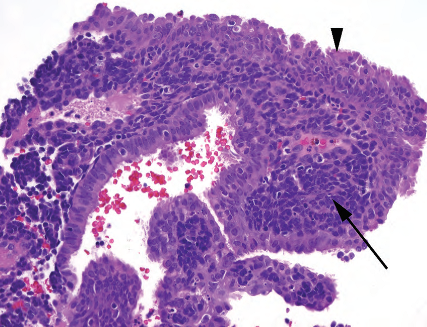
Figure 17.6. Endometrial stromal breakdown. The stroma is condensed into an extremely blue mass of tightly packed cells (arrow). The overlying epithelium is expanded into papillary tufts of pink cells, some with cilia, which is a metaplastic change (arrowhead).
图17.6 子宫内膜间质崩解。间质浓缩成非常蓝的紧密排列的细胞团(箭)。被覆的上皮伸展,形成粉红色的乳头状细胞丛,部分细胞有纤毛,这是一种化生改变(箭头)。
Endometritis: The diagnosis of acute endometritis requires microabscesses and epithelial destruction; the presence of neutrophils alone may just indicate menstrual breakdown. Chronic endometritis is diagnosed by the presence of plasma cells. In general, the stroma takes on a blue spindly look, and there are increased numbers of lymphocytes; these features should prompt you to crawl around at 20× looking for plasma cells (Figure 17.7).
子宫内膜炎:急性子宫内膜炎的诊断需要微脓肿和上皮破坏;单有中性粒细胞可能只是表明月经期崩解。慢性子宫内膜炎的诊断需要存在浆细胞。一般来说,间质呈蓝色梭形细胞,淋巴细胞增多;这些特点会提示你转到20倍并寻找浆细胞(图17.7)。
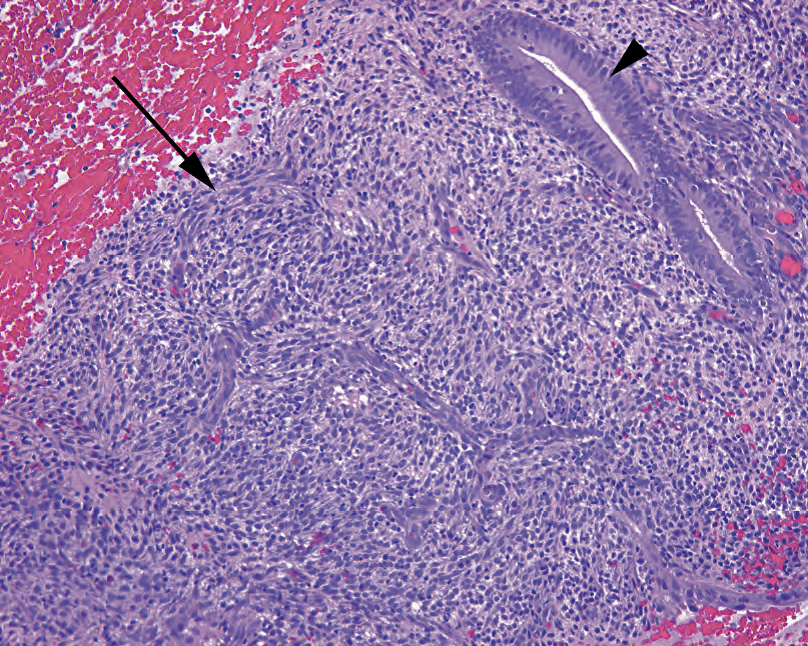
Figure 17.7. Chronic endometritis. At low power, the diagnostic plasma cells are not visible, but the spindly, swirling blue stroma (arrow) should be a clue to look more closely. The epithelium here is proliferative (arrowhead).
图17.7 慢性子宫内膜炎。低倍镜下看不到诊断性浆细胞,但梭形、漩涡状蓝色间质(箭)应该是诊断线索,促使你更仔细地观察。此处的上皮为增殖性(箭头)。
Disordered proliferative endometrium: Disordered proliferative endometrium is notable for a mixture of cystically dilated, budding, and tubular glands in a proliferative setting, with only focal glandular crowding. It occurs during anovulatory cycles.
紊乱的增殖性子宫内膜:主要表现为囊状扩张、出芽和管状腺体的混合,位于增殖性背景中,仅有局灶性腺体拥挤。它发生在无排卵周期。
Atrophy: Atrophy, described earlier, is responsible for about half of all cases of abnormal postmenopausal bleeding.
萎缩:如前所述,萎缩大约是绝经后异常出血的一半原因。
增生(Hyperplasia)
Hyperplasia is defined as an increase in the gland-to-stroma ratio, and you will notice it as “crowded glands” in a proliferative setting. Endometrial hyperplasia is categorized with two criteria: architecture (simple vs. complex) and cytology (with or without atypia). The term dysplasia is not applied to endometrium. There are varying degrees of hyperplasia:
增生定义为腺体与间质的比值增加,表现为增殖背景中的“拥挤的腺体”,从而引起你的注意。子宫内膜增生根据两种标准进行分类:结构(简单或复杂)和细胞学(有或无细胞异型性)。异型增生一词不适用于子宫内膜。有不同程度的增生:
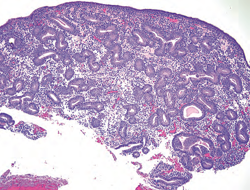
Figure 17.8. Simple hyperplasia. In this biopsy specimen, the glands appear proliferative and are too crowded (the gland-to-stroma ratio is greater than 1). The cells resemble normal endometrium and are not atypical.
图17.8 简单性增生。在该活检标本中,腺体增殖且过于拥挤(腺体与间质的比值大于1)。这些细胞与正常子宫内膜相似,并不非典型。
Simple hyperplasia: Simple hyperplasia almost always occurs without atypia. It appears as crowded, tubular, or minimally branched glands (Figure 17.8).
简单性增生:简单性增生几乎总是没有细胞异型性。它表现为拥挤的管状或轻微分支的腺体(图17.8)。(译注:简单性增生是指结构的简单)
Complex hyperplasia: Complex hyperplasia can occur with or without atypia. It appears as back-to-back glands with little stroma (even more crowded than in simple hyperplasia), and the glands have increasingly complex, branched outlines.
复杂性增生:可伴或不伴细胞异型性。它表现为背靠背的腺体,间质极少(远比单纯增生更密集),腺体具有更加复杂的分支状形状。(译注:复杂性增生是指结构的复杂)
Atypical hyperplasia: The definition of atypia varies among organs. In the endometrium, the normal proliferating gland has hyperchromatic, pseudostratified, elongated nuclei and frequent mitoses. This appearance in other organs, such as the colon, may make you think of low-grade dysplasia (such as a tubular adenoma). In endometrial atypia, the nuclei become round and pale or vesicular because of the chromatin clumping up and migrating to the nuclear membrane (Figure 17.9). Nucleoli may be prominent. The nuclei lose polarity and are seen at all levels of the epithelium (stratified). Nuclei are larger and show increased variability in size and shape. The cytoplasm becomes more eosinophilic than in nonatypical glands. Atypia is present or absent but is not graded.
非典型增生:细胞异型性的定义因器官而异。在子宫内膜中,正常增殖性腺体具有深染、假复层、拉长的细胞核、细胞核和频繁的核分裂。这种表现在其他器官,如结肠,可能使你想到低级别异型增生(如管状腺瘤)。子宫内膜的细胞异型性表现为核变圆、淡染或空泡状,这是由于染色质聚集成粗块状并迁移到核膜(图17.9)。核仁可能显著。细胞核失去极性,可见于上皮的各个层面(复层化)。细胞核增大,核大小和形状的变化程度更大。与正常腺体相比,其细胞质嗜酸性更强。诊断中只要说明异型性存在或不存在,不用分级。(译注:非典型增生是指具有细胞异型性,主要见于复杂性增生,此时称为复杂性非典型增生,见下文;极少见于简单性增生)
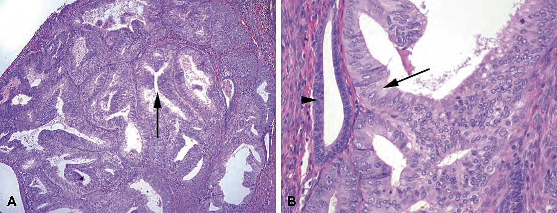
Figure 17.9. Complex atypical hyperplasia. (A) At low power, the glands are very crowded, even back to back, and the gland lumens have become branching and irregular (arrow). (B) At high power, comparing the hyperplastic epithelium (arrow) with normal residual glands (arrowhead), the hyperplastic cells have round nuclei, and pale, vesicular chromatin with prominent nucleoli, diagnostic of atypia.
图17.9 复杂性非典型增生。(A)在低倍镜下,腺体非常拥挤,甚至背靠背,腺腔变成分支状和不规则(箭头)。(B)在高倍镜下,比较增生上皮(箭)和正常残留腺体(箭头),增生细胞具有圆核和淡染、空泡状染色质,核仁显著,这些对细胞异型性具有诊断意义。
Complex atypical hyperplasia: Complex atypical hyperplasia (CAH) is a precursor to car-cinoma. Florid cases of CAH may in fact be hard to distinguish from well-differentiated endometrial carcinoma—so much so that experts may disagree. The concept of “carcinoma in situ” is not used in the endometrium, but CAH is fairly equivalent to it.
复杂性非典型增生(CAH):是癌前病变。实际上,CAH的旺炽性病例很难区分高分化子宫内膜癌,以至于专家们可能诊断不一致。子宫内膜不用“原位癌”的概念,但CAH相当于原位癌。
Do not be fooled by artifactual crowding in a biopsy. When glands are scraped out of the uterine cavity, they may clump together and look crowded. You need to find an intact piece of endometrium to evaluate the gland to stroma ratio. Also, beware of calling hyperplasia in the setting of an endometrial polyp (they are often crowded) or secretory endometrium.
不要被活检中人为假象造成的腺体拥挤所愚弄。当腺体被刮出宫腔时,它们可能会聚集在一起,看起来很拥挤。你需要找到一块完整的子宫内膜来评估腺体与间质的比值。也要注意,在子宫内膜息肉(腺体通常拥挤)或在分泌性子宫内膜的情况下,尽量不要称之为增生。
不孕症检查(The Infertility Workup)
When evaluating an endometrial biopsy specimen for infertility, first rule out the conditions listed earlier that can cause bleeding. Some infertility centers request that you date the endometrium, estimating the cycle day by histology. They are looking for a luteal phase defect, which is defined as a disparity of over 3 days between the calendar date and the histologic date in two consecutive biopsy specimens.
在评估不孕症的子宫内膜活检标本时,首先排除前面列出的可能导致出血的情况。一些不孕症中心要求你确定子宫内膜的日期,通过组织学来估计标本处于月经周期的第几天。他们正在寻找黄体期缺陷,黄体期缺陷定义为两个连续活检标本在日历上的实际日期和组织学估计日期之间的差异超过3天。
Proliferative endometrium cannot be dated. The first secretory change occurs, on average, on day 16 or so of a 28-day cycle. This change is the appearance of clear secretory vacuoles at the base of the epithelial cells, below the nuclei. When you see just a few of these in a generally proliferative endometrium, it is called interval endometrium. Beyond that day, specific histologic criteria are as follows:
增殖性子宫内膜不能确定日期。第一次分泌性改变平均发生在28天月经周期的第16天(译注:排卵后的第2天)左右。这种改变是在细胞核下方的上皮细胞底部出现透明的分泌空泡。在普遍增殖性子宫内膜中只看到少数核下空泡时,称为间期子宫内膜(译注:排卵后的第1天)。这一天以后,具体的组织学标准如下:
Days 16 to 20: glands are the most helpful feature
第16至20天:腺体是最有用的特征
Day 16: subnuclear vacuoles, pseudostratified nuclei
第16天:核下空泡,核假复层
Day 17: subnuclear vacuoles, but with an orderly row of nuclei (the “piano key” look)
第17天:核下空泡,核整齐排列(像是钢琴键那样)
Day 18: vacuoles above and below nuclei
第18天:核上和核下空泡
Day 19: few vacuoles, found only above nuclei; orderly row of nuclei, no mitoses
第19天:少数细胞含有核上空泡;细胞核排列整齐(译注:核回到基底部),无核分裂
Day 20: peak secretions in lumen and ragged luminal border, vacuoles rare
第20天:管腔分泌高峰,管腔边缘参差不齐,空泡罕见
From days 21 to 28, the glands change little. They are exhausted and appear low columnar with orderly nuclei, no mitoses, and ragged luminal edges. They may also have degenerative apical vacuoles—tricky to discern from days 19 to 20. After day 21, the stroma is the key:
从第21天到第28天,腺体改变不大。腺体分泌衰竭,呈低柱状,核整齐排列,无核分裂,管腔边缘参差不齐。在第19天到第20天,它们也可能有退化的顶端空泡,难以辨别。总之,第21天之后,间质是关键:
Day 21: beginning of stromal edema; secretion continues
第21天:间质水肿开始;分泌继续
Day 22: peak stromal edema with naked nuclei
第22天:间质水肿达到顶峰,(译注:间质细胞出现)裸核
Day 23: spiral arteries become prominent
第23天:螺旋动脉变得显著
Day 24: periarteriolar cuffing with predecidua (stromal cells around the arteries begin to get plump pink cytoplasm, creating a pink halo around the vessels)
第24天:动脉周围出现前蜕膜化(动脉周围的间质细胞开始形成丰满的粉红色细胞质,在血管周围形成粉红色的一圈)
Day 25: predecidual change under the surface epithelium
第25天:表面上皮的下方出现前蜕膜化
Day 26: decidual islands coalesce, lymphocytes begin to infiltrate stroma
第26天:蜕膜岛融合(译注:形成大范围前蜕膜化),淋巴细胞开始浸润间质
Day 27: many neutrophils in a solid sheet of decidua, with focal necrosis and hemorrhage 第27天:成片的实性蜕膜中出现许多中性粒细胞,伴局灶性坏死和出血
Day 28: prominent necrosis, hemorrhage, clumping, and break up
第28天:显著坏死、出血、结块和崩解
妊娠改变(Changes of Pregnancy)
Gestational endometrium is a solid sheet of decidua. Decidual cells are plump polygonal cells with pink to lavender cytoplasm and small oval nuclei. The glandular epithelium becomes almost papillary in nature with a hypersecretory appearance.
妊娠期子宫内膜是一层成片的实性蜕膜。蜕膜细胞为多角形细胞,胞质为粉红色至淡紫色,核小,卵圆形。腺上皮几乎变成乳头状,具有高分泌外观。
Well-formed glands with ballooning, cleared-out cytoplasm and wildly pleomorphic nuclei are characteristic of the Arias-Stella reaction, a normal reaction to pregnancy (Figure 17.10). The changes can be focal. The lack of mitoses or infiltration differentiates this from clear cell carcinoma, as does the age of the patient (clear cell is usually postmenopausal) and the surrounding gestational changes.
Arias-Stella反应是一种正常的妊娠反应,其特征是完好的腺体,腺体呈气球状膨胀,细胞质透明,广泛的核多形性(图17.10)。这种改变可是局灶性。缺乏核分裂或浸润,可区分透明细胞癌,患者年龄(透明细胞癌通常为绝经后)和周围妊娠改变也有助于鉴别。
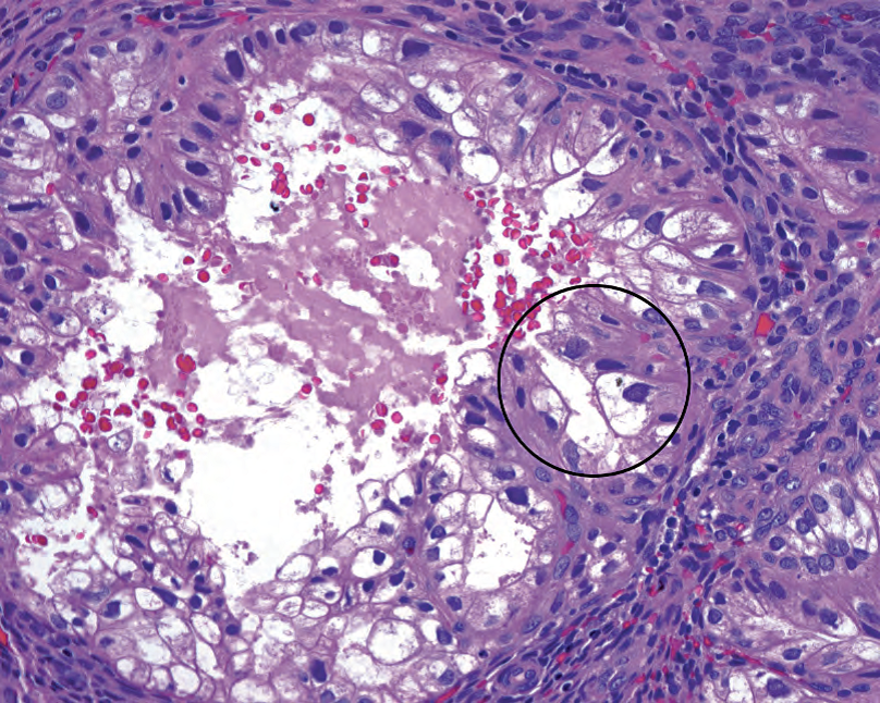
Figure 17.10. Arias-Stella reaction. Glands in the gestational endometrium can show bizarre cytology, including cleared-out cytoplasm and large hyperchromatic irregular nuclei (circle).
图17.10 Arias-Stella反应。妊娠期子宫内膜的腺体可能显示奇怪的细胞学,包括透明的细胞质,大的深染不规则核(圆圈)。
In a patient with a history of pregnancy, you may see a placental site nodule: aggregates of intermediate trophoblastic cells, which have scattered large nuclei that look atypical, within hyaline nodules. Placental site nodules should be well-circumscribed. They are the benign remnants of old implantation sites (see Figure 16.8 in Chapter 16).
有妊娠史的患者,你可能会看到胎盘部位结节:透明结节内的中间滋养层细胞聚集,具有散在的大核,貌似非典型核。胎盘部位结节应当界限清楚。它们是陈旧的胎盘种植部位的良性残余物(见第16章图16.8)。
化生类型(Types of Metaplasia)
Metaplasia by itself is a benign process; however, metaplasia is often accompanied by hyperplasia. Still, it is important to recognize these cell varieties and not call them cancer. The less ominous sounding word change may be used instead of metaplasia. Cell types include the following:
化生本身是良性过程;然而,化生常伴有增生。尽管如此,重要的是要认识到这些细胞种类,不要误诊为癌。听起来不那么可怕的名词“改变(change)”可以用来代替“化生(metaplasia)”。细胞类型包括以下几种:
Tubal metaplasia: luminal cilia in an epithelium that looks slightly plumped up and cleared out (If you overlook the cilia, the nuclei may appear atypical.)
输卵管化生:上皮的腺腔面出现纤毛,这些细胞看起来略微丰满和透明(如果你忽略了纤毛,会觉得核非典型)
Squamous metaplasia: swirling islands of immature squamous cells and, rarely, keratinization
鳞状化生:未成熟鳞状细胞形成旋涡状细胞岛,很少有角化
Mucinous metaplasia: mucinous, endocervical-type cells
粘液化生:粘液性,宫颈内膜型细胞
Eosinophilic metaplasia: increased eosinophilic cytoplasm; cells can proliferate in glands to the point of looking papillary (If the cells merge to form syncytial papillary tufts, it is papillary syncytial metaplasia.)
嗜酸性化生:嗜酸性细胞质增多;细胞可以在腺体中增殖,达到乳头状程度(如果细胞合并,形成合胞体乳头状细胞簇,称为乳头状合胞体化生)
Clear cell change: clear cells
透明细胞改变:透明细胞
子宫内膜恶性肿瘤(Endometrial Malignancies)
The endometrium has two cell types that can transform: glandular and stromal. Glandular cells give rise to several types of carcinoma, including endometrioid, serous, and clear cell. The stromal cells give rise to stromal sarcomas; these are entirely different from the leiomyosarcomas of the myometrium.
子宫内膜有两种细胞类型可能发生恶性转化:腺细胞和间质细胞。腺细胞形成几种类型的癌,包括子宫内膜样癌、浆液性癌和透明细胞癌。间质细胞形成子宫内膜间质肉瘤;这与子宫肌层来源的平滑肌肉瘤完全不同。
Endometrioid carcinoma is the most common type of endometrial cancer. It usually occurs in postmenopausal women (80% of cases), like its precursor lesion, complex atypical hyperplasia. Be cautious about diagnosing either type in a young woman, although it can happen.
子宫内膜样癌是最常见的子宫内膜癌类型。它通常发生在绝经后妇女(80%的病例),就像它的前驱病变——复杂性非典型增生(CAH),也是如此。在诊断年轻女性的癌或CAH时都要谨慎,尽管确实可能会发生。
Endometrioid carcinoma, in its well-differentiated form, closely resembles atypical endometrial glands. Architecturally, they are fused and complex and cover large areas without intervening stroma (Figure 17.11). The overall pattern may appear cribriform or villoglandular (like a villous adenoma of colon). The tumor may be limited to endometrium or may invade myometrium or adjacent organs; the extent determines stage. The grade is determined by cytology and architecture. High grade tumors are equivalent to “poorly differentiated.”
高分化的子宫内膜样癌很像非典型子宫内膜腺体。从结构上看,腺体融合而形成复杂结构,覆盖大片范围,腺体之间没有间质(图17.11)。整体模式可能呈筛状或绒毛状(就像结肠的绒毛状腺瘤)。肿瘤可能局限于子宫内膜,也可能浸润子宫肌层或邻近器官;浸润程度决定分期。肿瘤分级取决于细胞学和结构。高级别肿瘤相当于“低分化”。
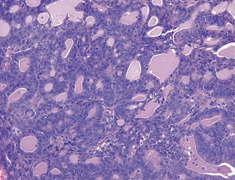
Figure 17.11. Endometrioid carcinoma. Foci of well-differentiated endometrioid carcinoma can be difficult to distinguish from complex atypical hyperplasia. However, the complicated proliferation of fused and cribriform glands in this biopsy specimen is diagnostic of carcinoma. The nuclei in this example resemble those of complex atypical hyperplasia.
图17.11 子宫内膜样癌。高分化子宫内膜样癌的病灶很难区分复杂性非典型增生。然而,在这例活检标本中,融合腺体和筛状腺体组成的复杂增殖已满足癌的诊断条件。本例细胞核类似于复杂性非典型增生。
FIGO (International Federation of Gynecology and Obstetrics) grade 1: The tumor is < 5% solid, where solid means sheets of cells that have lost their glandular differentiation. Areas of squamous metaplasia (common) are not counted as solid areas.
FIGO(国际妇产科联盟)1级:实性肿瘤<5%,其中实性指失去腺体分化的成片细胞。鳞状化生(常见)区域不算作实性区域。
FIGO grade 2: The tumor is 6%–50% solid.
FIGO 2级:实性肿瘤为6%-50%。
FIGO grade 3: The tumor is > 50% solid.
FIGO 3级:实性肿瘤>50%。
Significant nuclear atypia (one of those features that require experience to judge) can raise the grade by one level unless the tumor is already grade 3. Variants of endometrioid carcinoma include those with squamous differentiation, a villoglandular variant, a secretory variant, and a ciliated cell variant. These variants are identified only when the majority of the tumor takes on that morphology.
显著的核异型性(需要经验来判断的特征之一)可以将分级提高一级,除非肿瘤已经是3级。子宫内膜样癌的变异型包括鳞状分化型、绒毛腺型、分泌性和纤毛细胞型。只有当大多数肿瘤呈现上述形态时,才能称为某种变异型。
Serous carcinoma is a separate tumor pathway, leading to a distinct type of carcinoma. Serous carcinoma is not associated with hormonal exposure or endometrial hyperplasia. It is considerably more aggressive than endometrioid carcinoma and tends to be diagnosed in older women. It is, by definition, high grade and therefore is not graded.
浆液性癌是一种独立的肿瘤途径而导致的特殊类型癌。浆液性癌与激素暴露或子宫内膜增生无关。它比子宫内膜样癌更具侵袭性,并且往往发生于老年妇女。根据定义,它是高级别,因此不用分级。
Histologically, it resembles serous carcinoma of the ovaries (formerly known as papillary serous). Therefore, its hallmark is a papillary architecture, although this is not required for diagnosis. The papillae have broad or fine fibrovascular cores with complex branching (Figure 17.12). The cells are notable for extreme atypia, including cherry-red nucleoli, bizarre mitoses, and multinucleated cells. As in the ovary, psammoma bodies are common.
组织学上,它像卵巢浆液性癌(以前称为乳头状浆液性癌)。因此,它的特征是乳头状结构,但乳头不是诊断所必需。乳头具有或宽或细的纤维血管轴心,并有复杂的分支(图17.12)。细胞异型性极显著而闻名,包括樱桃红核仁、奇异的核分裂和多核细胞。与卵巢一样,砂粒体也很常见。
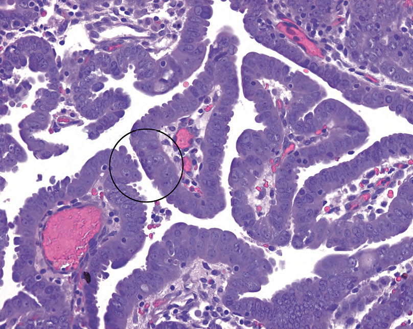
Figure 17.12. Serous carcinoma. Papillary structures are lined by atypical nuclei with prominent nucleoli (circle).
图17.12 浆液性癌。乳头状结构被覆着非典型细胞核,核仁显著(圆圈)。
The precursor lesion is believed to be endometrial intraepithelial carcinoma (EIC), a transformation of the surface epithelium, especially in polyps in older women. EIC is not quite analogous to carcinoma in situ, because EIC itself has metastatic potential. EIC, like serous carcinoma, is often associated with a p53 mutation leading to overexpression. An immunostain for p53 is sometimes used to confirm the diagnosis. Histologically, EIC appears as an abrupt transition on the surface from benign atrophic epithelium to pleomorphic, enlarged, atypical, mitotically active cells (Figure 17.13).
一般认为浆液性癌的前驱病变是子宫内膜上皮内癌(EIC)。EIC是表面上皮的一种转化,尤其是在老年妇女的息肉中。EIC并不等同于原位癌,因为EIC本身具有转移潜能。EIC与浆液性癌一样,常p53突变有关,p53突变导致过p53过表达,因此可用免疫染色检测,有时用于证实诊断。组织学上,EIC表现为从息肉表面的良性萎缩上皮到多形性、增大、非典型、核分裂活跃细胞的突然转变(图17.13)。
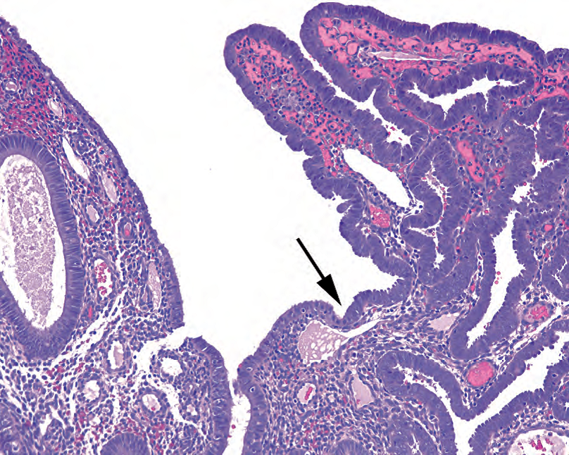
Figure 17.13. Endometrial intraepithelial carcinoma. There is an abrupt transition (arrow) from normal surface epithelium (left) to malignant cells (right). The cells of endometrial intraepithelial carcinoma resemble those of serous carcinoma.
图17.13 子宫内膜上皮内癌(EIC)。从正常表面上皮(左)到恶性细胞(右)的突然转变(箭)。EIC细胞类似于浆液性癌细胞。
Clear cell carcinoma, like serous carcinoma, occurs primarily in older women, has no relation to hormone exposure or hyperplasia, and has a poor prognosis. Histologically, it will remind you of clear cell neoplasms in other organs (such as renal cell). The cytoplasm is glycogen-rich and clear, the cell borders are distinct, and the architecture can be tubular, papillary, or solid. It may be mistaken for secretory endometrioid carcinoma but has much more nuclear pleomorphism. Like serous carcinoma, this tumor is high-grade by definition. Other rare types of carcinoma include squamous, mucinous, transitional cell, undifferentiated, and small cell carcinoma.
透明细胞癌和浆液性癌一样,主要发生于老年妇女,与激素暴露或增生无关,预后差。组织学上,它会让你想起其他器官(如肾)的透明细胞肿瘤。细胞质富含糖原,透明,细胞边界清晰,结构可以是管状、乳头状或实性。可能误认为分泌性子宫内膜样癌,但核多形性更明显。与浆液性癌一样,这种肿瘤定义为高级别。其他罕见类型癌包括鳞癌、粘液癌、移行细胞癌、未分化癌和小细胞癌。
Endometrial stromal sarcoma is a rare malignancy of the endometrial stromal cells. The difference between a benign stromal nodule and a low-grade endometrial stromal sarcoma is in the interface with the surrounding tissue—sarcomas are infiltrative. These sarcomas have minimal atypia and few mitoses, as well as a prominent plexiform vascular proliferation (like the normal spiral arteries gone wild). The high-grade endometrial stromal sarcoma, however, has marked atypia and bizarre mitoses, like most high-grade sarcomas.
子宫内膜间质肉瘤是一种罕见的子宫内膜间质细胞恶性肿瘤。良性间质结节和低级别子宫内膜间质肉瘤的区别在于与周围组织的交界处,肉瘤是浸润性的。这些肉瘤的异型性很小,核分裂很少,并有明显的丛状血管增殖(就像正常的螺旋动脉,失控生长)。然而,高级别子宫内膜间质肉瘤与大多数高级别肉瘤一样,具有明显的异型性和奇异的核分裂。
Malignant mullerian mixed tumor (carcinosarcoma) is a mixed tumor consisting of malignant glands in a sarcomatous stroma. It appears as a recognizable carcinoma, such as endometrioid type, with adjacent sarcomatous cells (large angular pleomorphic nuclei) in the stroma (Figure 17.14). Other soft tissue elements, like skeletal muscle or cartilage, may also show up. It is similar in concept to a carcinosarcoma of other organs.
恶性苗勒混合瘤(癌肉瘤,MMMT)是一种混合性肿瘤,由肉瘤间质中的恶性腺体组成。它表现为一种可识别的癌类型(如子宫内膜样癌),间质中有相邻的肉瘤细胞(大的成角的多形性核)(图17.14)。其他软组织成分,如骨骼肌或软骨,也可能出现。它的概念类似于其他器官的癌肉瘤。
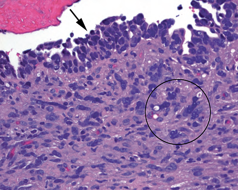
Figure 17.14. Malignant mullerian mixed tumor. This tumor is defined by the presence of carcinomatous cells in the epithelium (arrow) and sarcomatous cells in the stroma (circle). The carcinomatous cells are hyperchromatic and crowded; elsewhere in this biopsy specimen there were malignant glands. The sarcomatous cells are hyperchromatic, large, and irregular in shape, similar to malignant fibrous histiocytoma–type cells found in other sarcomas (see Chapter 28).
图17.14 恶性苗勒混合瘤(癌肉瘤,MMMT)。定义为上皮中存在癌细胞(箭)并且间质中存在肉瘤细胞(圆圈)。癌细胞深染拥挤;这例活检标本的其他部位有恶性腺体。肉瘤细胞呈深染的大而不规则形状,类似于其他肉瘤中发现的恶性纤维组织细胞瘤型细胞(见第28章)。
In contrast, an adenosarcoma is a neoplasm with benign glands and a malignant stroma. An adenofibroma is benign glands with benign stroma. These tumors are similar to the phyllodes tumor of the breast, which can range from malignant to benign stroma.
相比之下,腺肉瘤是一种良性腺体和恶性间质组成的混合性肿瘤。腺纤维瘤兼有良性腺体和良性间质。这两种肿瘤类似于乳腺叶状肿瘤,其间质包括从恶性到良性范围。
子宫肌层(Myometrium)
The most common neoplasm of the uterus is actually the leiomyoma, a benign smooth muscle tumor of the myometrium. These tumors can be huge, multiple, myxoid, even necrotic, and still benign. Although benign, many are removed for symptomatic relief. When sampling these at the grossing bench, what you are looking for are areas that are different in texture from the typical rubbery dense consistency; areas of necrosis, hemorrhage, or dense white foci should be sampled.
子宫最常见的肿瘤实际上是平滑肌瘤,一种子宫肌层的良性平滑肌肿瘤。这些肿瘤可以是巨大的、多发的、黏液样、甚至坏死,并且仍然是良性的。虽然是良性,但许多是为了缓解症状而切除。取材时,你要寻找质地不同于典型橡胶样致密程度的区域;坏死、出血或致密白色病灶区域都要取材。
The classic leiomyoma is a spindle cell lesion with intersecting fascicles of elongated cells, typically intersecting at right angles (Figure 17.15). The nuclei are long and thin with fine pale chromatin and small nucleoli. You may also see “corkscrew” nuclei, which are characteristic of smooth muscle. The stroma may be fibrotic, edematous, myxoid, or even hemorrhagic; these are all permissible degenerative changes in the absence of nuclear atypia or a high mitotic rate.
典型的平滑肌瘤是一种梭形细胞病变,由拉长的细胞束交叉而成,通常呈直角交叉(图17.15)。细胞核细长,染色质细腻淡染,核仁小。你也可以看到“拧毛巾样”细胞核,这是平滑肌的特征。间质可能纤维化、水肿、粘液样,甚至出血;在没有核异型性或高核分裂率的情况下,这些改变都是允许的退行性改变。
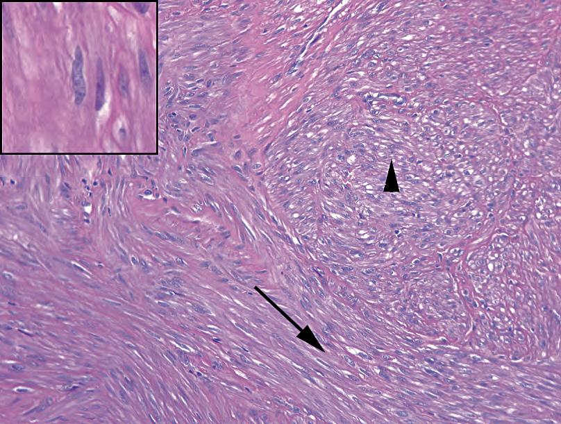
Figure 17.15. Leiomyoma. The low-power impression is that of fascicles or bundles of cells, some parallel to the slide (arrow) and some coming out at right angles (arrowhead). Inset: The nuclei are tapered and pale, with occasional paranuclear vacuoles, and sometimes show “corkscrew” morphology, as though the nucleus was twisted longitudinally. (Dog owners may liken this lumpy shape to something else.)
图17.15 平滑肌瘤。低倍印象是细胞束,有些平行于切片(箭),有些呈直角(箭头)。插图:细胞核一端渐细(译注:一般形容平滑肌核“两端钝圆”),淡染,偶有核旁空泡,有时呈“拧毛巾样”形态,似乎细胞核被纵向扭曲。(养狗的人可能会把这种块状的形状比作其他东西。)(译注:直白说“狗屎”)
Leiomyosarcoma tends to present as a large, solitary mass and is not thought to arise from preexisting leiomyomas. It may resemble the fascicular leiomyoma, but mitotic activity must be high, over 10 per 10 high-power fields, and cytologic atypia should be prominent (Figure 17.16). (In sarcomas, atypia takes the form of large dark nuclei with crisp, irregularly shaped nuclear borders.) The third feature is coagulative necrosis. The threshold for diagnosing leiomyosarcoma is quite high in the uterus, unlike a leiomyomatous lesion found in the soft tissue or retroperitoneum, for example. In the uterus, atypia without mitotic activity, or mitoses without atypia, should discourage you from calling a sarcoma.
平滑肌肉瘤往往表现为一个巨大的孤立性肿块,一般认为不是来自先前存在的平滑肌瘤。它可能类似于束状平滑肌瘤,但核分裂活性必须很高,每10个高倍视野中有10个以上核分裂象,并且细胞异型性应当显著(图17.16)。(在肉瘤中,细胞异型性表现为核大而深染,核边界清晰而不规则。)第三个特征是凝固性坏死。子宫平滑肌肉瘤的诊断门槛很高,不同于软组织或腹膜后发现的平滑肌肿瘤。在子宫中,异型性不伴核分裂活跃,或核分裂活跃不伴异型性,不要称为肉瘤。
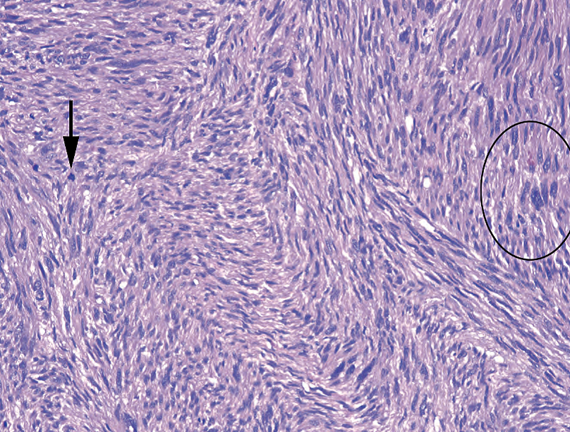
Figure 17.16. Leiomyosarcoma. The threshold for diagnosing leiomyosarcoma in the uterus is high. This lesion should be a much more cellular version of the leiomyoma, with mitoses (arrow), atypical and pleomorphic cells (circle), and necrosis (not seen here).
图17.16 平滑肌肉瘤。诊断子宫平滑肌肉瘤的门槛很高。这种病变应该远比平滑肌瘤的细胞更丰富,伴有核分裂(箭头)、非典型和多形性细胞(圆圈)以及坏死(此处未看到)。
Adenomatoid tumor is a benign proliferation of mesothelial origin. It often occurs on the serosal surface of the uterus, resembling a leiomyoma both grossly and microscopically. The mesothelial tumor cells induce a smooth muscle proliferation that is probably often mistaken for leiomyoma. However, on close inspection, you will see small clefted spaces between the muscle bundles, lined by cuboidal cells forming gland-like or angiomatoid lumens. The cells can appear epithelial by histology and in fact would stain for cytokeratins. Accidentally missing it and calling it a leiomyoma does no harm to the patient; mistaking it for metastatic adenocarcinoma would be disastrous, however. Unlike adenocarcinoma, it should stain for calretinin.
腺瘤样瘤是间皮起源的良性增殖。它通常发生在子宫浆膜面,在大体和显微镜下都像平滑肌瘤。间皮肿瘤细胞诱导平滑肌增殖,可能容易误诊为平滑肌瘤。然而,仔细观察,你会看到肌束之间的小的裂隙状空隙,被覆立方细胞,形成腺样或血管瘤样空腔。这些细胞在组织学上表现为上皮样细胞,事实上,CK阳性。不小心漏诊,称为平滑肌瘤对患者没有伤害;然而,误诊为转移性腺癌将是灾难性的。与腺癌不同,它应该表达calretinin。
(译注:腺瘤样瘤取材时有粘滑感;补充图片如下)
来源:
The Practice of Surgical Pathology:A Beginner’s Guide to the Diagnostic Process
外科病理学实践:诊断过程的初学者指南
Diana Weedman Molavi, MD, PhD
Sinai Hospital, Baltimore, Maryland
ISBN: 978-0-387-74485-8 e-ISBN: 978-0-387-74486-5
Library of Congress Control Number: 2007932936
© 2008 Springer Science+Business Media, LLC
仅供学习交流,不得用于其他任何途径。如有侵权,请联系删除。
本站欢迎原创文章投稿,来稿一经采用稿酬从优,投稿邮箱tougao@ipathology.com.cn
相关阅读
 数据加载中
数据加载中
我要评论

热点导读
-

淋巴瘤诊断中CD30检测那些事(五)
强子 华夏病理2022-06-02 -

【以例学病】肺结节状淋巴组织增生
华夏病理 华夏病理2022-05-31 -

这不是演习-一例穿刺活检的艰难诊断路
强子 华夏病理2022-05-26 -

黏液性血性胸水一例技术处理及诊断经验分享
华夏病理 华夏病理2022-05-25 -

中老年女性,怎么突发喘气困难?低度恶性纤维/肌纤维母细胞性肉瘤一例
华夏病理 华夏病理2022-05-07


共0条评论