外科病理学实践:诊断过程的初学者指南 | 第15章 卵巢(Ovary)
第15章 卵巢(Ovary)
The ovary is a fairly straightforward organ as far as the pathologist is concerned. Find tumor, remove tumor, identify tumor. No messing around with nonneoplastic pathology, reactive lesions, and so forth. If it looks malignant, it probably is.
对病理医生来说,卵巢是相当简单的器官。发现肿瘤,切除肿瘤,识别肿瘤。不要乱搞非肿瘤性病理学、反应性病变等。如果它看起来是恶性的,很可能就是恶性。
正常组织学和定义(Normal Histology and Definitions)
Surface epithelium: Surface epithelium is essentially a mesothelial lining. It is easily rubbed off the surface, so you do not always see it. Most epithelial tumors (the most common neoplasms) are thought to arise from this epithelium or from invaginations of it. Think of it as a pluripotent stem cell layer.
表面上皮:表面上皮实质上是被覆的间皮。它很容易从表面脱落,所以你并不是总能看到它。一般认为大多数上皮性肿瘤(也是最常见的卵巢肿瘤)起源于表面上皮或其内陷。把它想象成多能干细胞层。
Stroma: The ovarian stroma is blue and spindly, with a streamy, fascicular look. Most of the cells in the stroma are fibroblasts (Figure 15.1).
间质:卵巢间质呈蓝色,梭形,呈束状排列。间质中的大多数细胞是成纤维细胞(或译作纤维母细胞)(图15.1)。
Sex cord cells: Sex cord cells are the hormone-secreting supporting cells of the ovary, the thecal cells and granulosa cells. The thecal cells, under luteinizing hormone stimulation, secrete androgens, and the granulosa cells, under follicle-stimulating hormone control, convert androgens to estrogen. Together they nurture an oocyte to ovulation.
性索细胞:性索细胞是卵巢分泌激素的支持性细胞,即卵泡膜细胞和粒层细胞。在促黄体生成素刺激下,卵泡膜细胞分泌雄激素,而在卵泡刺激素控制下,粒层细胞将雄激素转化为雌激素。它们一起培育卵母细胞以排卵。
Follicles: The follicles are characterized by a halo of thecal cells outside a ring of granulosa cells (see Figure 15.1), all surrounding the giant oocyte (germ cell). In developing follicles, the granulosa cells form Call-Exner bodies, rosettes of granulosa cells surrounding pink globules.
卵泡(或译作滤泡):卵泡的特征是在环形排列的粒层细胞之外还有一圈卵泡膜细胞(见图15.1),所有这些细胞都围绕着巨大的卵母细胞(生殖细胞)。在发育中的卵泡中,粒层细胞形成Call-Exner小体,这是粒层细胞围绕粉红色小体而形成的花环结构。
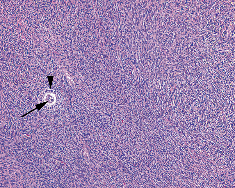
Figure 15.1. Ovarian stroma with follicle. Typical ovarian stroma is blue and cellular, with a vaguely fascicular or storiform pattern. A small primary follicle is seen with the central oocyte (arrow) and a ring of granulosa cells (arrowhead).
图15.1.卵巢间质,含有卵泡。典型的卵巢间质呈蓝色,细胞丰富,呈模糊的束状或席纹状。一个小的初级卵泡,中央有卵母细胞(箭)和一圈粒层细胞(箭头)。
Luteinized: Similar to decidualized, luteinized indicates cells that have become plump with abundant pink cytoplasm.
黄素化:与蜕膜化类似,黄素化表示细胞变得丰满,具有丰富的粉红色细胞质。
Corpus luteum: The corpus luteum is a newly ovulated follicle (Figure 15.2). The capsule of luteinized granulosa cells collapses in on itself, becoming undulating, and there is associated hemorrhage. The corpus luteum produces progesterone until (and if) the placenta takes over. If there is no pregnancy, it involutes.
黄体:黄体是一个新的排卵后卵泡(图15.2)。黄素化粒层细胞组成的包膜自身塌陷,变得上下起伏,伴有出血。黄体产生黄体酮,直到(如果怀孕)胎盘接管。如果没有怀孕,黄体就会退化。
(译注,上文将围绕卵细胞的多层黄素化粒层细胞视为卵细胞的包膜,并不是真包膜)
Corpus albicans: The former corpus luteum ultimately hyalinizes to form cloud-shaped pink islands in the ovary, scars of old follicles (see Figure 15.2).
白体:先前黄体最终透明变性,在卵巢中形成云状粉红色岛,旧卵泡形成瘢痕(见图15.2)。
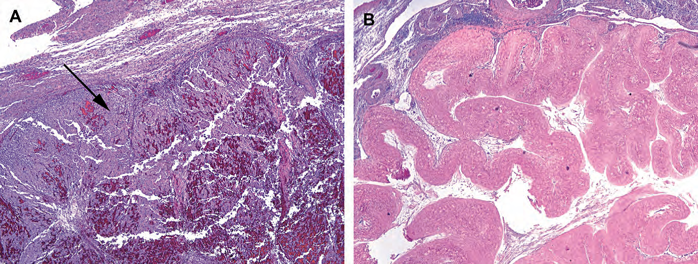
Figure 15.2. (A) Hemorrhagic corpus luteum, with undulating layers of luteinized granulosa cells (arrow) and associated blood. (B) A corpus albicans, the remnant of a prior corpus luteum.
图15.2.(A) 出血性黄体,具有上下起伏的黄素化粒层细胞(箭),伴有出血。(B) 白体是先前黄体的残余物。
Walthard’s rests: Walthard’s rests are nests of transitional (urothelial) type epithelium in the ovary and fallopian tube.
Walthard细胞巢:是卵巢和输卵管中的移行细胞(尿路上皮)形成的细胞巢。
Rete ovarii: Analogous to the rete testis in men, rete ovarii are rudimentary gland spaces located in the hilum of the ovary. They are angulated, slit-like spaces with a low cuboidal epithelium (Figure 15.3). Do not mistake them for cancer.
卵巢网:类似于男性的睾丸网,卵巢网是位于卵巢门的未充分发育的腺腔。它们是成角的狭长空隙,具有低立方形上皮(图15.3)。不要误认为是癌症。
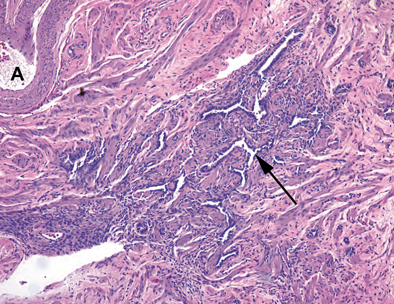
Figure 15.3. Rete ovarii. This vestigial structure is found at the hilum of the ovary, adjacent to large arteries (A) and veins. The rete consist of slit-like channels with a cuboidal cell lining (arrow).
图15.3.卵巢网。这种退化结构位于卵巢门,毗邻大动脉(A)和静脉。卵巢网衬覆立方形细胞,形成狭长空隙(箭)。
Follicle cyst: A follicle cyst is lined with the normal components of the follicle, the granulosa cells, and the thecal layer (Figure 15.4). A similar lesion is the hemorrhagic corpus luteum cyst, which is a blood-filled corpus luteum.
卵泡囊肿:卵泡囊肿衬覆卵泡的正常成分,即粒层细胞和卵泡膜细胞组成(图15.4)。类似的病变是出血性黄体囊肿,是一种充满血液的黄体。
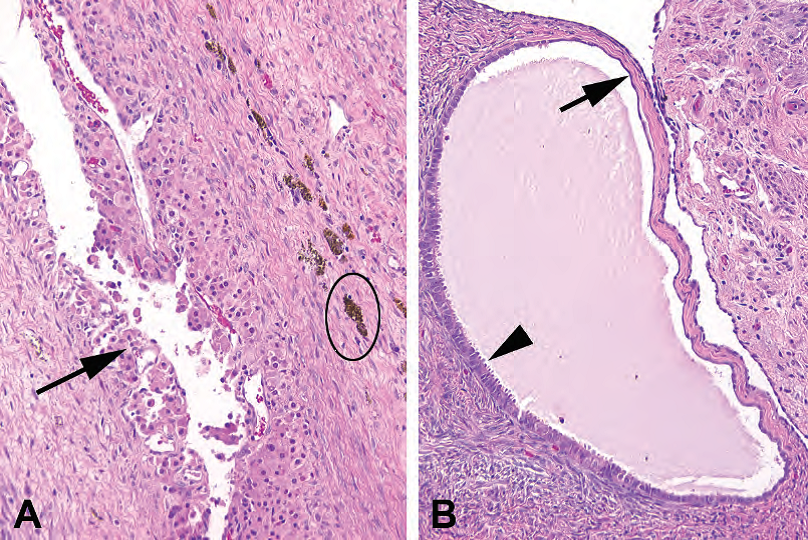
Figure 15.4. Follicular cyst versus inclusion cyst. (A) A follicle cyst is lined by luteinized cells, similar to those seen in the corpus luteum (arrow). There is adjacent hemosiderin (oval). (B) An inclusion cyst may be lined by an attenuated epithelium, similar to the surface epithelium (arrow), or may show tubal metaplasia (arrowhead).
图15.4.卵泡囊肿与包涵囊肿的对比。(A) 卵泡囊肿衬覆黄素化细胞(箭),类似于黄体。旁边有含铁血黄素(椭圆形)。(B) 包涵囊肿可能衬覆变薄的上皮,类似于表面上皮(箭头),也可能显示输卵管化生(箭头)。
Inclusion cyst: An inclusion cyst is a simple cyst lined with a cuboidal, columnar, or ciliated epithelium, often budding inward from the ovarian surface (see Figure 15.4). When small, these can be called surface inclusion cysts. However, if they are large, they are best referred to as serous cystadenomas (see later).
包涵囊肿:是一种简单的囊肿,内衬立方、柱状或纤毛上皮,通常从卵巢表面向内出芽(见图15.4)。总体积小的时候,可以称为表面包涵囊肿。然而,如果巨大,最好称为浆液性囊腺瘤(见下文)。
肿瘤(Neoplasms)
For each cell type defined above (and then some), there are families of neoplasms that can arise. Table 15.1 lists the types of neoplasms that can occur. Those in parentheses are rare enough that we will not talk about them here.
上面定义的每种细胞类型(以及其中一部分),都可能发生一组肿瘤。表15.1列举可能发生的肿瘤类型。括号中的肿瘤非常罕见,我们在此不讨论。
Table 15.1. Neoplasms of the ovary.
表15.1.卵巢肿瘤。
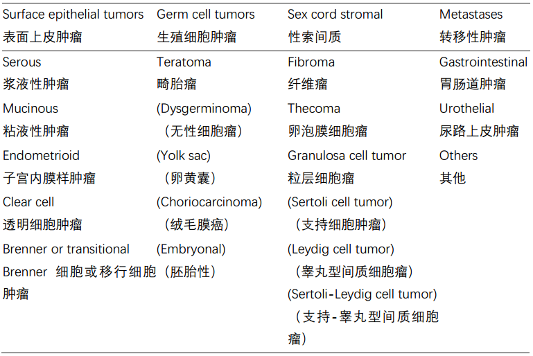
Note: Entries in parentheses are rare tumors that are not discussed in this chapter.
注:括号中的内容是本章未讨论的罕见肿瘤。
上皮性肿瘤(Epithelial Neoplasms)
Epithelial neoplasms are one of three types: benign, borderline, and malignant. Benign tumors do not metastasize, malignant ones do, and borderline tumors may recur or rarely metastasize. The nomenclature is as follows:
上皮性肿瘤有三种类型:良性、交界性和恶性。良性肿瘤不转移,恶性肿瘤转移,交界性肿瘤可能复发或很少转移。命名法如下:
Adenoma: An adenoma is a benign epithelial proliferation. When cystic, as they often are, it is a cystadenoma. If biphasic with a secondary fibrous stromal component, it is an adenofibroma. If all three, it is a cystadenofibroma. Histologically, the cystadenomas are simple or multilocular cysts with a flat epithelial lining.
腺瘤:腺瘤属于良性上皮增殖性病变。通常囊性,称为囊腺瘤。如果是双相性,即,既有囊肿,又有继发性纤维间质成分,称为腺纤维瘤。如果这三种情况都存在,那就是囊腺纤维瘤。组织学上,囊腺瘤是简单的囊肿或多房性囊肿,衬覆扁平上皮。
Borderline tumor (atypical proliferative tumor, low malignant potential): Borderline lesions have an increasing epithelial complexity over the adenomas. Their epithelium begins to ruffle up in papillary fronds and may “ruffle down” into the stroma in a way that looks similar to invasion. However, they do not cross the basement membrane, do not invade as single cells, and do not induce a desmoplastic reaction. Borderline tumors can shed cells into the peritoneum, which may stick onto other organs and begin to grow like weeds. However, they often do not technically invade and are called noninvasive implants, not metastases. Invasive implants can also occur and act like true metastases.
交界性肿瘤(非典型增殖性肿瘤,低度恶性潜能):与腺瘤相比,交界性病变的上皮更加复杂。上皮开始向上皱折成乳头状结构,也可能向下皱折进入间质,这种生长方式貌似浸润。然而,它们不穿透基底膜,没有单个细胞浸润,也不诱导促结缔组织增生反应。交界性肿瘤可以将细胞脱落到腹膜中,腹膜可能粘附在其他器官上,并开始像杂草一样生长。然而,从原理上讲,它们通常既不是浸润,也不是转移瘤,而是称为非浸润性种植。浸润性种植也可能发生,并表现得像是真正的转移。
Carcinoma: Carcinomas commonly present as combination cystic/solid tumors and are called cystadenocarcinomas. However, there is not a significant clinical difference between calling something a carcinoma and a cystadenocarcinoma. These can be divided into low- and high-grade carcinomas, but all types can metastasize.
癌:通常表现为囊性肿瘤/实性肿瘤,称为囊腺癌。然而,将它们称为癌和囊腺癌之间并没有显著的临床差异。这些肿瘤可分为低级别和高级别癌,但所有类型均可转移。
Carcinosarcoma: A carcinosarcoma (malignant mixed mullerian tumors in the gynecologic tract) is a carcinoma in a sarcomatous stroma. An adenosarcoma would be a benign epithelial neoplasm in a sarcomatous stroma, which is rare.
癌肉瘤:癌肉瘤(女性生殖的恶性苗勒混合瘤,MMMT)是肉瘤性间质中的癌。腺肉瘤是肉瘤性间质中的良性上皮性肿瘤,腺肉瘤很少见。
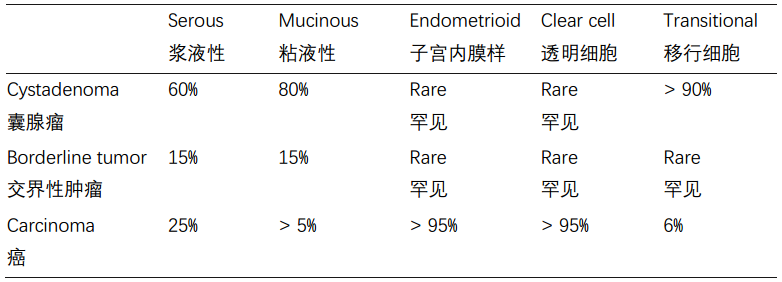
Within the surface epithelial group, there are five types of epithelial neoplasms. Each type can be subdivided into benign, borderline, or malignant, as shown in Table 15.2.
在表面上皮组中,有五种类型的上皮肿瘤。每种类型可以细分为良性、交界性或恶性,如表15.2所示。
Table 15.2. Five types of epithelial neoplasms.
表15.2.五种上皮性肿瘤。
浆液性(最常见,以前称为乳头状浆液性)(Serous (Most Common, Formerly Papillary Serous))
Cystadenoma: Cystadenomas are simple cysts lined by a tubal-like epithelium with columnar and/or ciliated cells. They can become huge, but the lining remains simple. The contents are watery.
囊腺瘤:囊腺瘤是一种简单的囊肿,衬覆输卵管样上皮,含有柱状和/或纤毛细胞。它们可以变得巨大,但衬覆上皮仍然简单。囊内容物是水样物。
Borderline tumor (atypical proliferative tumor; tumor of low malignant potential): Borderline tumors have increasingly complex papillary fronds, looking grossly granular. The papillary pattern is characterized by tree-like branching of smaller and smaller papillae (Figure 15.5). However, the epithelium lining the papillae is usually a single layer without significant atypia. When the papillae acquire secondary epithelial proliferations called micropapillae (the medusahead look; Figure 15.6), they are on their way to micropapillary serous carcinoma (MPSC). A few millimeters of confluent micropapillary pattern upgrades this to an actual MPSC, in some textbooks.
交界性肿瘤(非典型增殖性肿瘤;低度恶性潜能肿瘤):交界性肿瘤具有复杂的乳头状结构,肉眼看似颗粒状。乳头状结构的特征是越来越小的树枝状乳头(图15.5)。然而,乳头被覆的上皮通常是单层,没有明显的异型性。当乳头变成微乳头,获得继发性上皮增殖时(像是美杜莎的头发,蛇发怪样结构;图15.6),它们开始向微乳头状浆液性癌(MPSC)进展。在一些教科书中,几毫米的融合性微乳头模式就会升级为实际的微乳头状浆液性癌。
Micropapillary serous carcinoma (micropapillary borderline tumor): Micropapillary serous carcinoma is a low-grade carcinoma characterized by the medusa-head pattern (see Figure 15.6). The nuclei should not be too pleomorphic. This carcinoma can occur within a confined cyst (noninvasive MPSC) or break out of the cyst and into the stroma (invasive). This perplexing concept of a noninvasive carcinoma is somewhat like the papillary urothelial carcinomas of the bladder or the papillary carcinomas of breast. Psammoma bodies are common. When invasive, the tumor nests have a flower-like shape, with nuclei pointing outwards, and often sit in small cleft-like spaces (Figure 15.6)
微乳头状浆液性癌(微乳头状交界性肿瘤):微乳头状浆液性癌是一种低级别癌,特征是美杜莎头发模式(见图15.6)。核多形性不太明显。这种癌可以发生在局限性囊肿内(非浸润性MPSC),也可以从囊肿破裂并进入间质(浸润性)。非浸润性癌这种令人困惑的的概念有点像膀胱的乳头状尿路上皮癌或乳腺的乳头状癌。砂粒体常见。发生浸润时,肿瘤巢呈小花形状,细胞核向外,通常位于小的狭长空隙内(图15.6)
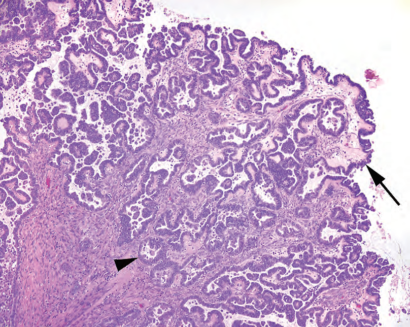
Figure 15.5. Borderline serous tumor. The epithelial lining is composed of serous, or nonmucinous, cells (arrow). The overall architecture is quite complex, with papillary branching and invaginated folds that should not be mistaken for invasion (arrowhead). However, the epithelial component is mostly a monolayer.
图15.5.交界性浆液性肿瘤。被覆上皮为浆液性,或非粘液性细胞(箭)。整体结构相当复杂,有乳头状分支,也有内陷的皱折,不要误认为浸润(箭头)。然而,上皮成分主要是单层。
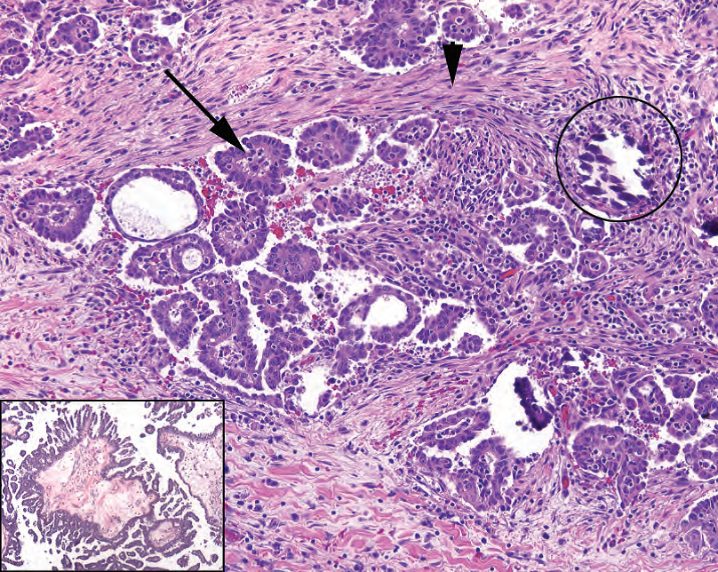
Figure 15.6. Micropapillary serous carcinoma. When invasive, micropapillary serous carcinoma looks like tiny florets of cells (arrow) in a desmoplastic stroma (arrowhead). Psammoma bodies are common (circle). Inset: The medusa-head, or micropapillary, pattern is indicative of micropapillary serous carcinoma. Compare the epithelial micropapillae to the simple epithelium of the borderline tumor (see Figure 15.5).
图15.6.微乳头状浆液性癌。发生浸润时,微乳头状浆液性癌看似小花细胞(箭),位于促结缔组织增生性间质(箭头)中。砂粒体常见(圆圈)。插图:美杜莎头发模式,或微乳头状结构,提示微乳头状浆液性癌。比较上皮性微乳头与交界性肿瘤的简单上皮(见图15.5)。
High-grade serous carcinoma: High-grade serous carcinoma has very high-grade, mitotically active, apoptotic, pleomorphic blue nuclei (Figure 15.7). The architecture can be papillary, micropapillary, solid, or in nests with slit-like spaces.
高级别浆液性癌:核级别非常高、核分裂活跃、凋亡、多形性蓝核(图15.7)。结构可以是乳头状、微乳头状、实性,也可以是具有狭长空隙的巢状结构。
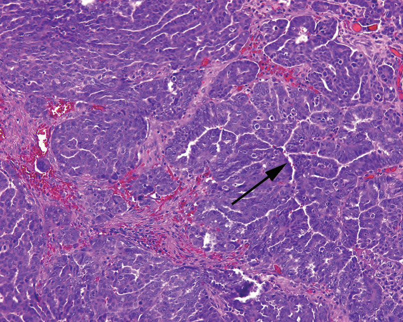
Figure 15.7. High-grade serous carcinoma. This tumor is shown invading the stroma. The cells are pleomorphic and dark, with prominent nucleoli, and grow in solid nests with slit-like spaces (arrow).
图15.7.高级别浆液性癌。图示肿瘤浸润间质。细胞呈多形性,深染,核仁显著,形成实性癌巢,位于狭长空隙(箭)中。
粘液性(Mucinous)
Cystadenoma: Cysts (often multilocular) are lined with fairly flat mucinous epithelium.
囊腺瘤:囊肿(通常多房性)内衬相当平坦的粘液性上皮。
Borderline tumor (atypical proliferative tumor; tumor of low malignant potential): The vast majority of these are of the intestinal type, which means they imitate intestinal epithelium, with goblet cells and glandular architecture. However, 15% are of the endocervical type, which presents as papillary architecture (like serous) with low-grade, squared-off, endocervical-like mucinous cells (Figure 15.8).
交界性肿瘤(非典型增殖性肿瘤;低度恶性潜能肿瘤):绝大多数为肠型,很像肠上皮,具有杯状细胞和腺体结构。然而,15%为宫颈内膜型,表现为乳头状结构(像浆液性肿瘤),伴有低级别、长方形、宫颈内膜样粘液细胞(图15.8)。
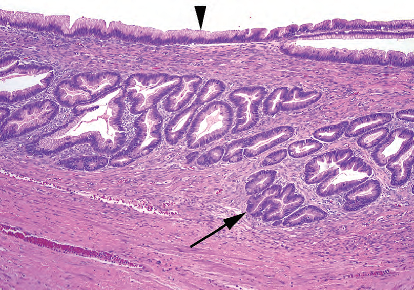
Figure 15.8. Borderline mucinous tumor. The cyst lining (arrowhead) is mucinous and resembles endocervical cells in this example. As with the borderline serous tumor, invaginations into the stroma should not be mistaken for invasion (arrow).
图15.8.交界性粘液性肿瘤。囊肿被覆黏液性上皮(箭头),本例类似宫颈内膜细胞。与交界性浆液性肿瘤一样,上皮成分内陷进入间质,不要误认为浸润(箭)。
Cystadenocarcinoma: Mucinous cystadenocarcinoma is very uncommon. Mucinous carcinoma in the ovary is usually a metastasis from the gastrointestinal tract.
囊腺癌:黏液性囊腺癌非常少见。卵巢粘液癌通常是胃肠道转移。
子宫内膜样(Endometrioid)
Adenoma: How is endometrioid adenoma different from endometriosis? It has no endometrial stroma. Also, these adenomas are rare. Endometriosis is common.
腺瘤:子宫内膜样腺瘤与子宫内膜异位症有何不同?它没有子宫内膜间质。此外,这些腺瘤罕见,而子宫内膜异位症常见。
Carcinoma: Endometrioid carcinomas imitate endometrial carcinoma and so have similar architecture to the patterns found in the uterus, including tubular to cribriform glands and villous structures (Figure 15.9). They may arise within endometriosis, they are often found along with endometriosis, and a concurrent endometrial carcinoma is not uncommon. They may be low or high grade.
癌:子宫内膜样癌类似(子宫的)子宫内膜癌,因此具有类似结构,包括管状至筛状腺体和绒毛状结构(图15.9)。它们可能发生在子宫内膜异位症中,通常与子宫内膜异位症一起发现,并发子宫内膜癌并不少见。它们可能是低级别,也可能高级别。
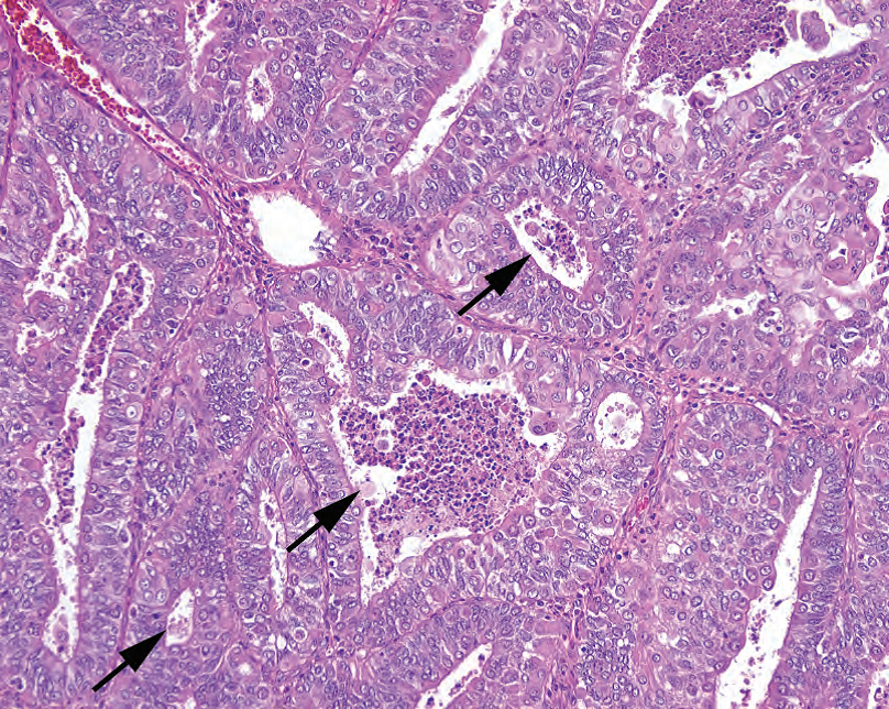
Figure 15.9. Endometrioid carcinoma. The nuclei are cleared out and pleomorphic, like endometrioid carcinoma of the endometrium. Distinct glandular spaces are visible (arrows), some with central necrosis.
图15.9.子宫内膜样癌。细胞核非常醒目,多形性,就像子宫内膜的子宫内膜样癌。可见明显的腺腔(箭),一些腺腔有中央坏死。
透明细胞(Clear Cell)
Carcinoma: Clear cell carcinomas are clear cells occurring in papillary, glandular, nested, or trabecular patterns. Cells tend to fall out of the center of the nests, leaving a hobnailed layer of cells outlining the nest (Figure 15.10). These are high grade and, like endometrioid carcinoma, are also associated with endometriosis.
细胞学表现为透明细胞,结构表现为乳头状、腺样、巢状或小梁状模式。细胞倾向于从巢的中心脱落,留下一层靯钉样细胞来,勾勒出巢的轮廓(图15.10)。透明细胞癌是高级别肿瘤,与子宫内膜样癌一样,也与子宫内膜异位症有关。
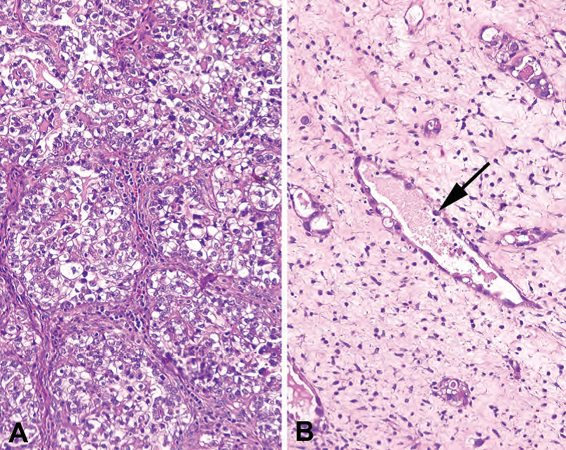
Figure 15.10. Clear cell carcinoma. (A) In this field, nests of clear cells are seen separated by fibrovascular septa. (B) A less cellular area of the same tumor shows vessel-like spaces lined by atypical cells that protrude into the lumen in hobnail fashion (arrow).
图15.10.透明细胞癌。(A) 在这个区域,可见透明细胞巢被纤维血管间隔分开。(B) 同一肿瘤细胞较少的区域,显示血管样腔隙,衬覆非典型细胞,呈鞋钉样突向腺腔(箭)。
移行细胞(Transitional)
Adenoma/adenofibroma: Transitional adenomas/adenofibromas are the Brenner tumor, characterized by nests of transitional epithelium in a fibrous stroma (Figure 15.11). There may be a mucinous layer surrounding a central lumen in each nest.
腺瘤/腺纤维瘤:移行性腺瘤/腺纤维瘤是Brenner肿瘤,其特征是纤维间质中的移行上皮巢(图15.11)。每个巢的中央管腔周围可能有一层粘液性细胞。
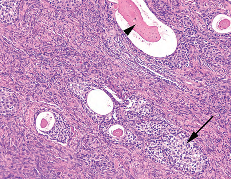
Figure 15.11. Brenner tumor. Nests of transitional-type epithelium (resembling urothelium; arrow) in a fibrotic stroma are typical. Some form gland-like spaces with pink secretions (arrowhead).
图15.11.Brenner肿瘤。典型表现为纤维化间质中的移行细胞型上皮巢(类似于尿路上皮;箭)。有些形成腺样腔隙,有粉红色分泌物(箭头)。
Malignant Brenner tumors: Malignant Brenner tumors are characterized by very atypical cells. Transitional cell carcinoma (resembling that seen in the bladder) is a term used when there is no coexisting Brenner element.
恶性Brenner肿瘤:恶性Brenner肿瘤的特征是细胞异型性非常明显。没有共存Brenner成分时,称为移行细胞癌(类似于膀胱癌)。
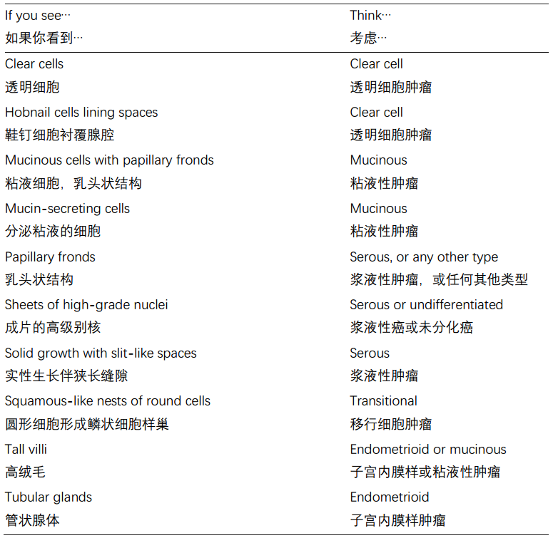
How can you tell which pattern you have?
你怎么能分辨你看到了哪种模式?
癌症途径(Cancer Pathways)
There are thought to be two cancer pathways for serous neoplasms:
浆液性肿瘤有两种癌症途径:
Cystadenoma → Atypical proliferative serous tumors → Micropapillary serous carcinoma → Invasive micropapillary serous carcinoma
囊腺瘤→ 非典型增殖性浆液性肿瘤→ 微乳头状浆液性癌→ 浸润性微乳头状浆液性癌
Terribly bad luck → High-grade serous carcinoma (de novo), often p53 positive
倒霉透了→ 高级别浆液性癌(从头发生,译注:没有癌前病变),通常p53阳性
非上皮性肿瘤(Nonepithelial Neoplasms)
For the purposes of this chapter, we will gloss over the germ cell and sex cord stromal neoplasms, except for the most common entities.
在本章中,我们将对生殖细胞和性索间质肿瘤进行概述,只讨论最常见的肿瘤实体。
生殖细胞肿瘤(Germ Cell Neoplasms)
Anything that can occur in the testis can occur in the ovary. The most common germ cell neoplasm in the ovary is the teratoma, but you can also see dysgerminoma (seminoma), yolk sac tumor, choriocarcinoma (arising unrelated to gestational trophoblastic disease), and embryonal carcinoma. Each looks similar in testis and ovary.
睾丸可能发生的任何东西,卵巢也可以。卵巢最常见的生殖细胞肿瘤是畸胎瘤,但也可以看到无性细胞瘤(精原细胞瘤)、卵黄囊瘤、绒毛膜癌(与妊娠滋养细胞疾病无关)和胚胎性癌。它们在睾丸和卵巢中都相似。
Teratomas are usually composed of at least two of three embryonic derivatives: ectoderm, endoderm, and mesodermal cells. They are often cystic (dermoid cyst) and may grow to a large size. Common elements include squamous epithelium, skin adnexal structures, hair, fat, cartilage, thyroid, brain and nerve tissue, gut epithelium, and respiratory epithelium. Primary nonovarian neoplasms can arise in teratomas, creating an endless list of case reports. Teratomas restricted to mature elements are benign in the ovary.
畸胎瘤通常至少由三种胚胎衍生物中的两种组成:外胚层、内胚层和中胚层细胞。它们通常是囊性(皮样囊肿),可能会长得很大。常见成分包括鳞状上皮、皮肤附件结构、毛发、脂肪、软骨、甲状腺、脑和神经组织、肠上皮和呼吸上皮。畸胎瘤中可能发生原发性非卵巢肿瘤,病例报告不胜枚举。卵巢中,局限于成熟成分的畸胎瘤是良性的。
All teratomas must be carefully evaluated for immature (embryonal-looking) elements. The most common immature tissue type is brain. After a few products-of-conception (POC) specimens or fetal autopsies, you should recognize fetal brain—dark blue cells in a myxoid, clear background (Figure 15.12).
所有畸胎瘤必须仔细评估是否存在未成熟(胚胎样)成分。最常见的未成熟组织类型是大脑。观察一些妊娠产物(POC)标本或胎儿尸检之后,你应该能识别胎儿大脑—黏液样透明背景中的深蓝色细胞(图15.12)。
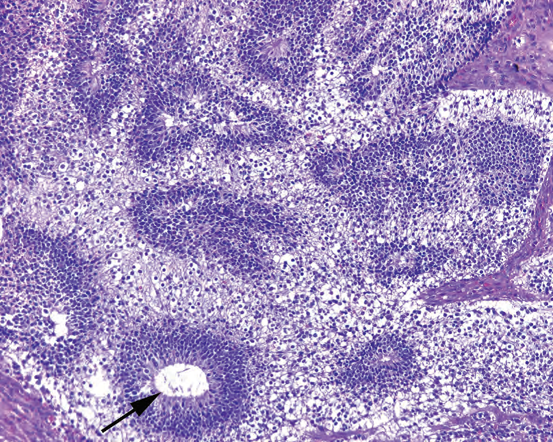
Figure 15.12. Immature neural tissue, teratoma. The combination of hypocellular areas and dense small round blue cell areas are suggestive of fetal brain. Rosettes (arrow) may also be seen. Finding this histology in a teratoma indicates an immature component.
图15.12.未成熟神经组织,畸胎瘤。少细胞区和密集的小圆蓝色细胞区的组合提示胎儿大脑。也可以看到菊形团(箭)。在畸胎瘤中发现这种组织学提示不成熟成分。
性索间质肿瘤(Sex Cord Stromal Neoplasms)
Sex cord stromal neoplasms include all of the fibrous and sex hormone cell types. The most common tumors are the fibroma/thecoma group. Granulosa cell tumors are also not uncommon. However, many of the weird and paradoxical ovarian lesions (Sertoli-Leydig?) fall into this group. Cell types in these tumors may be luteinized, just like their normal counterparts.
性索间质肿瘤包括所有的纤维细胞和性激素细胞类型。最常见的肿瘤是纤维瘤/卵泡膜瘤组。粒层细胞瘤也不少见。然而,许多奇怪和矛盾的卵巢病变(Sertoli-Leydig?)也属于性索间质肿瘤。这些肿瘤中的细胞类型可能黄素化,就像它们的正常对应物一样。
The fibroma/thecoma group is a spectrum of lesions from the pure fibroma, to the common mixed fibrothecoma, to the pure thecoma. Grossly they look like leiomyomas, which are very rare in the ovary. On cross section, the thecoma areas are butter-colored, and stand out from the grey-white fibroma. Histologically the tumors are also similar to leiomyomas but have more of a sheet-like pattern with bland, spindled cells (Figure 15.13). However, the tiny lipid vacuoles that identify the thecoma component (steroid cells, remember?) are very hard to see on H&E stain, so the gold standard is an oil red O done on frozen section. Bright red lipid globules indicate a thecoma component. These are benign tumors.
纤维瘤/膜细胞瘤组是一个病变谱系,从单纯的纤维瘤到普通的混合性纤维卵泡膜细胞瘤再到单纯的卵泡膜细胞瘤。大体上,它们看起来像平滑肌瘤,而平滑肌瘤在卵巢中非常罕见。横切面上,卵泡膜瘤区域呈奶油色,与灰白色纤维瘤明显不同。组织学上,肿瘤也类似于平滑肌瘤,但更多表现为成片的形态学温和的梭形细胞(图15.13)。然而,在HE染色切片上,识别卵泡膜成分(类固醇细胞,记得吗?)的微小脂质空泡很难看到,因此金标准是冷冻切片上的油红O染色。亮红色的脂质球表明卵泡膜成分。这些是良性肿瘤。
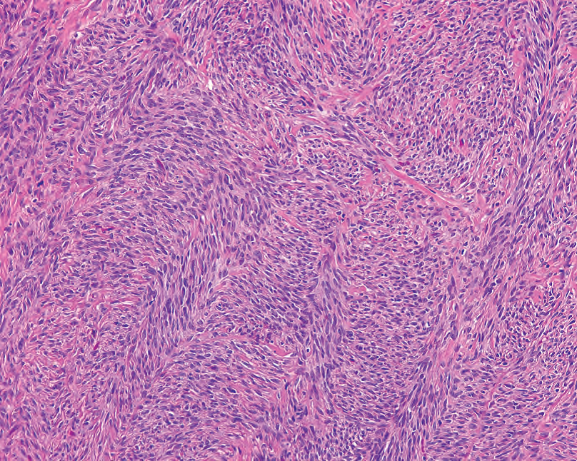
Figure 15.13. Fibrothecoma. This specimen shows mainly the fibroma component, with fascicles of bland spindled cells.
图15.13.纤维膜瘤。该标本主要是纤维瘤成分,形态学温和的梭形细胞呈束状排列。
Granulosa cell tumor cells appear similar to the normal granulosa cells in the ovary but have more distinctive oval folded or angulated nuclei with a longitudinal groove (the “coffee bean” nuclei). In a tumor, these cells may become more closely packed, almost giving the impression of nuclear molding, but they are not as blue, hyperchromatic, or crowded as small cell carcinoma. At low power the cells are arranged in sheets, with a zigzag “watered silk” pattern (think of a topographic map; Figure 15.14). The cells appear very uniform throughout. Rarely, you may see the pathognomonic Call-Exner bodies as seen in the developing follicle. These are technically of low malignant potential but may recur after many years.
粒层细胞肿瘤细胞与卵巢中的正常粒层细胞相似,但具有更独特的卵圆形折叠的或成角的细胞核,并有纵向核沟(“咖啡豆样”核)。在肿瘤中,这些细胞可能变得更加紧密,几乎形成核成型的印象,但它们不像小细胞癌那样蓝色、深染或密集。在低倍镜下,细胞成片排列,具有锯齿形的“水绸样”模式(想象一下地形图;图15.14)。总体上,细胞形态看起来非常均匀一致。可能偶尔见到具有病理诊断意义的Call-Exner小体,正如在发育中的卵泡中所见。从技术上讲,这些肿瘤是低度恶性潜能,但多年后可能复发。
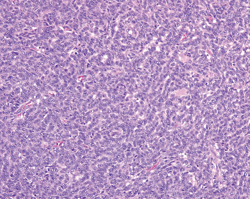
Figure 15.14. Granulosa cell tumor, low power. This section shows the characteristic cords and rows of granulosa cells, creating a pattern like watered silk, or (for those not frequenting fabric stores), a topographic map.
图15.14.粒层细胞瘤,低倍镜。这一部分显示粒层细胞的特征性条索和成行排列,形成了一种类似于水绸的模式,或(对于那些不经常光顾织物商店的人)像是地形图。
来源:
The Practice of Surgical Pathology:A Beginner’s Guide to the Diagnostic Process
外科病理学实践:诊断过程的初学者指南
Diana Weedman Molavi, MD, PhD
Sinai Hospital, Baltimore, Maryland
ISBN: 978-0-387-74485-8 e-ISBN: 978-0-387-74486-5
Library of Congress Control Number: 2007932936
© 2008 Springer Science+Business Media, LLC
仅供学习交流,不得用于其他任何途径。如有侵权,请联系删除。
本站欢迎原创文章投稿,来稿一经采用稿酬从优,投稿邮箱tougao@ipathology.com.cn
相关阅读
 数据加载中
数据加载中
我要评论

热点导读
-

淋巴瘤诊断中CD30检测那些事(五)
强子 华夏病理2022-06-02 -

【以例学病】肺结节状淋巴组织增生
华夏病理 华夏病理2022-05-31 -

这不是演习-一例穿刺活检的艰难诊断路
强子 华夏病理2022-05-26 -

黏液性血性胸水一例技术处理及诊断经验分享
华夏病理 华夏病理2022-05-25 -

中老年女性,怎么突发喘气困难?低度恶性纤维/肌纤维母细胞性肉瘤一例
华夏病理 华夏病理2022-05-07


共0条评论